
Presentation
Recurrent UTI, diagnosed on US basis as left pelviureteric junction obstruction and referred to MR urography prior to pyeloplasty.
Patient Data


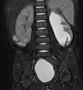
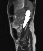

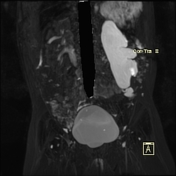
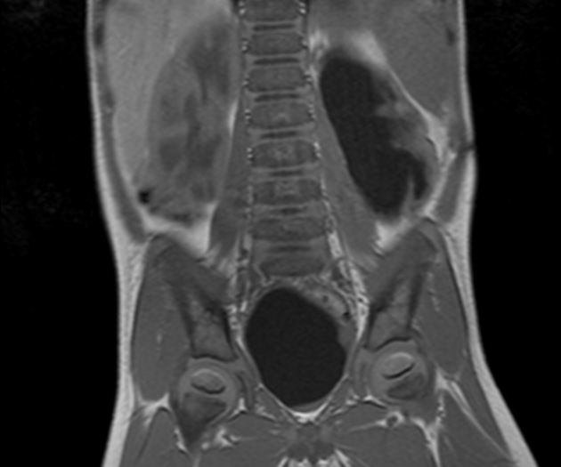
The left kidney is average in size measuring 80 x 23 x 25 mm. Renal Parenchyma showed decreased thickness (4 mm), cortical scars, normal signal and poor corticomedullary differentiation. Moderate central and peripheral calyceal dilatation. The renal Pelvis is moderately dilated with AP diameter: 19 mm. The dilatation showed abrupt termination at the pelviureteric junction. The left ureter is not dilated.
Case Discussion
This case demonstrates typical appearances of left PUJ obstruction, likely congenital. Congenital pelviureteric junction obstruction is commonly unilateral, more common in males and it is by far the most common cause of pediatric hydronephrosis. It is demonstrated as dilatation of pelvicalyceal system with abrupt end at pelviureteric junction. the ureter is usually not dilated and this differentiates it from vesicoureteric reflux disease.


 Unable to process the form. Check for errors and try again.