Presentation
Reported history of congestive heart failure with worsening shortness of breath.
Patient Data
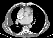

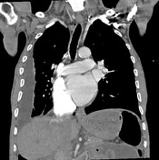

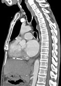

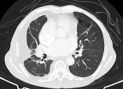

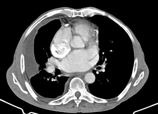
Right pleural thickening with small loculated effusion. Partially imaged lobulated abdominal ascites.
Pericardial thickening and scattered calcifications. Distortion of the normal heart contours with elongation of the ventricles and compression of the atria. Dilation of the right atrium and intrahepatic segment of the IVC.
Mild centrilobular emphysema. Interstitial edema and likely some degree of rounded atelectasis in the right lung.
Case Discussion
Distortion of the normal cardiac contour indicates constrictive physiology due to thickening and calcifications of the pericardium. This causes interstitial edema in the right lung.
The presence of right pleural thickening with effusion and partially imaged peritoneal thickening/ascites indicates an infectious process such as TB (endemic in the patient's area) as the cause.


 Unable to process the form. Check for errors and try again.