
Presentation
Anterior ischemia on ECG.
Patient Data


CTCA
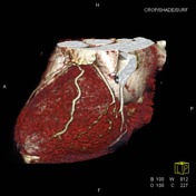
Coronary arteries:
Origins: Normal although the RCA arises from between the right and non coronary sinuses.
Dominance: Right
Left Main Coronary Artery (LMCA): Normal.
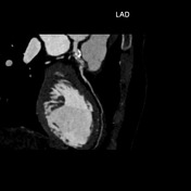
Left Anterior Descending (LAD): fusiform aneurysm in the proximal segment which is difficult to adequately assess given the artefact. It is irregular, 17 mm long and 6 mm in diameter with mural calcification proximally. Mild non opacification in the proximal portion of the aneurysm is suspicious for thrombosis.
D1, D2 - Patent
Circumflex artery (Cx): Normal.
1st obtuse marginal branch (OM1) - Patent
Intermediate artery (Ix): large vessel, normal.
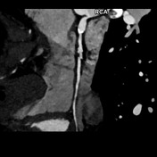
Right Coronary Artery (RCA): 2 fusiform aneurysms without thrombosis- 1) proximal segment 15 mm long and 8 mm in diameter with mural calcification and 2) mid segment 7 mm long and 6.5 mm in diameter at the origin of a small AM branch.
Posterior descending artery (PDA) - Patent
Posterior left ventricular branch (PLV) - Patent
Cardiovascular Findings:
No LV myocardial thinning or fatty metaplasia.
Other Findings
Incidental 1.5cm segment 8 arterially enhancing lesion.
Conclusion
LAD and RCA aneurysms. No stenotic disease evident.
Case Discussion
Further Hx is of Kawasaki disease since 3 months of age. Anterior infarct from LAD thrombosis at 8 months.


 Unable to process the form. Check for errors and try again.