
Patient Data
- Note: This case has been tagged as "legacy" as it no longer meets image preparation and/or other case publication guidelines.

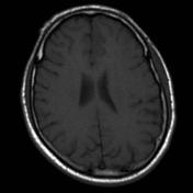

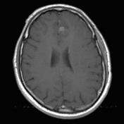
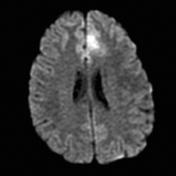
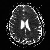
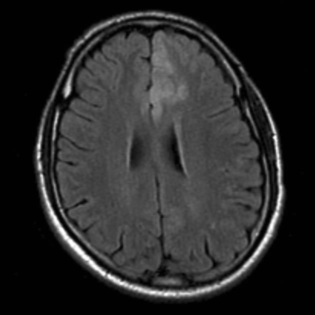
There is a region of increased T2 signal with positive mass effect involving the superior frontal gyrus. A faint region of contrast enhancement is present.
Case Discussion
Features are consistent with a diffuse low-grade glioma. Only faint enhancement and absence of evidence of necrosis/hemorrhage argues against a higher grade lesion (i.e unlikely to be a GBM).
Histology
Paraffin sections show fragments of white matter. These show a moderate increase in the numbers of astrocytic cells and prominent perineuronal and perivascular secondary structuring. Astrocytic cells show a mild degree of nuclear atypia and there is clustering of cells in many areas. No mitotic figures are seen and there is no vascular endothelial cell proliferation and no necrosis. A minority of atypical astrocytic cells show strong nuclear staining for Ki-67/MIB-1 (proliferation index approx. 5%). The features are of diffuse fibrillary astrocytoma.
FINAL DIAGNOSIS: diffuse (fibrillary) astrocytoma
Note: IDH mutation status is not provided in this case and according to the current (2016) WHO classification of CNS tumors, this tumor would, therefore, be designated as a diffuse astrocytoma NOS.


 Unable to process the form. Check for errors and try again.