
Presentation
Mild SOB post endovascular closure of patent foramen ovale (PFO).
Patient Data

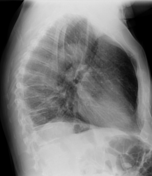
The PFO closure device is projected over the upper abdomen in the posteriorly, just to the left of midline suggesting it is in the aorta rather than the IVC.
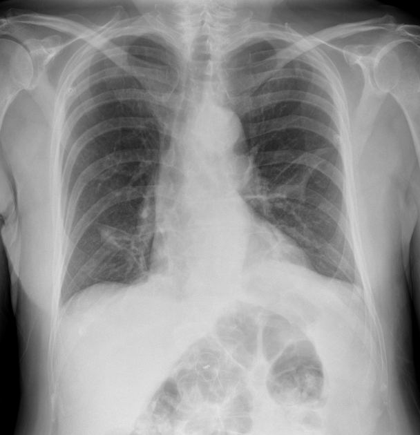
Extensive areas of atelectasis have developed in the left mid and both lower zones with elevation of the left hemidiaphragm. There is minor blunting of both CP angles.
The cardiomediastinal contours are unchanged.
During the procedure, echo showed the device was in the correct position within the PFO but slightly loose even after re-sizing.
The device was then retrieved the endovascularly from the aorta and the next day a different size device was deployed.
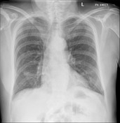
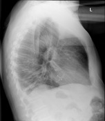

The PFO closure device now appears correctly positioned.
Persisting bilateral lower lobe areas of patchy collapse. The left mid zone atelectasis has resolved. Small right pleural effusion.
Case Discussion
Always worth checking any device for its position and integrity.


 Unable to process the form. Check for errors and try again.