
Presentation
Dyspnea, myalgias, cough, frequent emesis, and generalized abdominal pain.
Patient Data
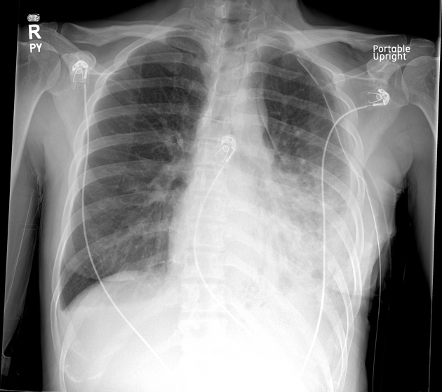
There is a small medial left pneumomediastinum. An apically oriented left chest tube is in place. There are airspace opacities in the left mid-lower lung. There are small bilateral pleural effusions.
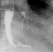
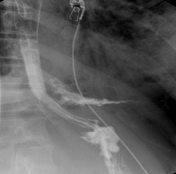

Frontal views of an esophagram shows prompt extravasation of contrast extending into the left lateral thorax.
Case Discussion
This is a case of an esophageal perforation. The patient was taken to the operating room emergently and she was found to have a left-sided empyema. She underwent an EGD with stent placement and a left video-assisted thoracoscopic decortication. She was admitted to the surgery intensive care unit and her postoperative course was unremarkable.


 Unable to process the form. Check for errors and try again.