
Presentation
Hypertension work-up.
Patient Data
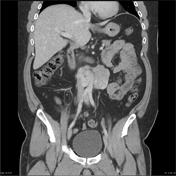
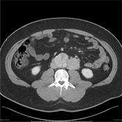

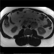


Mass in the region of the organ of Zuckerkandl measures 10 x 4 x 7 cm ( craniocaudal ). This lesion is only of moderate patchy T2 hyperintensity. Postcontrast images delineate the lower abdominal aorta separated from the mass by a thin rim of non-or hypoenhancing tissue. There is mass-effect on the inferior vena cava, but no obvious invasion. Heavy T2-weighted fat-suppressed sequence concurs - the IVC and aortic wall integrity appears preserved.
Conclusion: Appearances are consistent with two discrete large extra-adrenal pheochromocytomas (paragangliomas).
Case Discussion
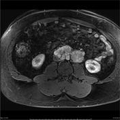
The masses encase and compress the IVC and aorta, which lie between the two lesions. The aortic and caval walls are well seen on T2 imaging and appear intact with no MR evidence of vascular invasion (given the extent of compression and circumferential involvement microscopic invasion can not be excluded). Both lesions are partly cystic and appear to be encapsulated, favoring a benign cause. Whilst the inferior mesenteric artery is completely encased for the proximal 10 mm or so of its course, the encapsulation suggests that it is also likely free of direct tumor invasion.


 Unable to process the form. Check for errors and try again.