
Presentation
Incidental finding during work-up for abdominal pain.
Patient Data
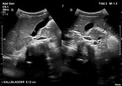
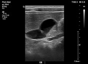
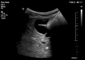
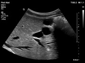

A partially distended gallbladder in a non-fasting patient (child). There are numerous gallbladder folds present. The gallbladder is otherwise normal.
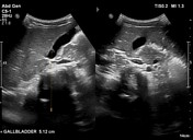
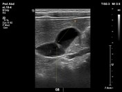
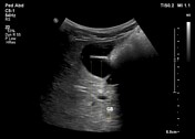
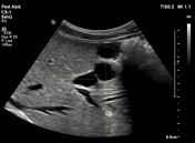
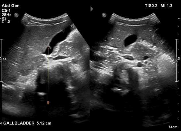
Arrows point to the gallbladder folds identified during the ultrasound examination. There are no well-defined gallstones, no sludge, normal wall thickness, and no pericholecystic fluid present.
Case Discussion
Gallbladder folds are usually thick, junctional in nature, and non-continuous or incomplete. A non-distended gallbladder or gallbladder in non-fasting patient may account for the wall folding on itself and giving rise to the particular sonographic appearance. Gallbladder septa are usually thin and often complete. They may be seen with gallbladder polyps and comet tail artifacts in adenomyomatosis.


 Unable to process the form. Check for errors and try again.