
Presentation
Infant with cough & breathlessness.
Patient Data
Age: 2 years
Gender: Male
Note: This case has been tagged as "legacy" as it no longer meets image preparation and/or other case publication guidelines.
From the case:
Ganglioneuroma

Download
Info
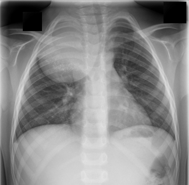
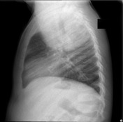
There is a voluminous, well-defined round lesion, located in the posterior mediastinum. There are powderish calcifications at the lower third of this lesion. Note the significant positive mass effect on the trachea and right main bronchus.
From the case:
Ganglioneuroma
Download
Info
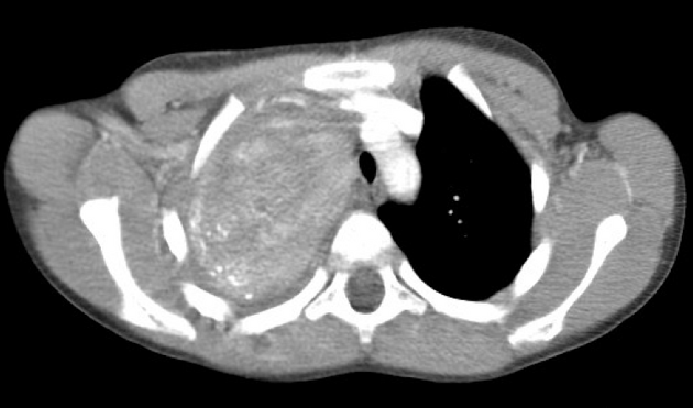
Single axial image from the CT chest with contrast, confirming the large posterior mediastinal lesion, with foci of calcification.
Case Discussion
Final pathologic diagnosis: ganglioneuroma.


 Unable to process the form. Check for errors and try again.