
Presentation
Weight loss.
Patient Data
Age: 50 years
Gender: Male
From the case:
Gastric adenocarcinoma


Download
Info

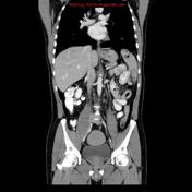
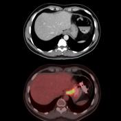
Abdomen CT demonstrates a subtle circumferential wall thickening involving the gastric cardia and gastroesophageal junction. High FDG uptake is noted at the gastric cardia confirming the presence of the tumor.
Download
Info
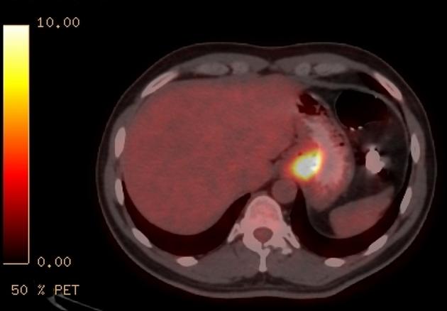
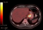
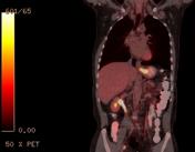
High FDG uptake is noted at the gastric cardia confirming the presence of the tumor.
Case Discussion
CT demonstrates a subtle circumferential wall thickening involving the gastric cardia and gastroesophageal junction. High FDG uptake is noted at the gastric cardia confirming the presence of a malignant tumor.
Pathology:
Moderately differentiated gastric cardiac adenocarcinoma. Distal esophagus is normal.


 Unable to process the form. Check for errors and try again.