
Presentation
A few weeks history of abdominal distention, bloating, malaise and fatigue. Mildly elevated liver function tests. Referred for an abdominal ultrasound.
Patient Data
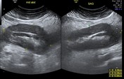

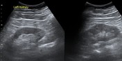

Diffuse circumferential mural thickening noted of the gastric wall, measuring at least 2.1 cm - pseudokidney appearance of the stomach. Adjacent surrounding fat stranding and pelvic free fluid.
Advised for upper GI endoscopy and staging CT.
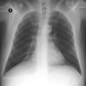
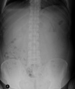

Absent gastric air bubble noted on the PA chest radiograph.
Apparent thickened gastric mucosa with slit like configuration (not reliable).

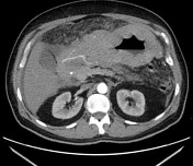
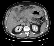
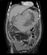
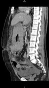
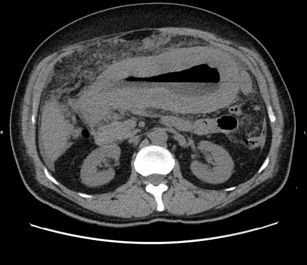
Diffuse heterogeneous circumferential gastric mural thickening (over 2 cm thick), extending into the first and second parts of the duodenum. Nodular appearance of mucosa suggesting submucosal infiltration. A few hypodense lesions are noted in the gastric wall representing necrosis/cystic changes. There is an exophytic area noted on the left side side of the gastric body. Findings typical for linitis plastica.
Extension along the gastrohepatic ligament and also along the gastrocolic ligament associated with mild thickening of part of the transverse colon.
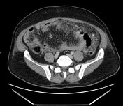
Extensive peritoneal involvement with thickening and enhancement. Involvement of the greater omentum and small bowel mesentery which enhances extensively. Loss of fat plane along the head and neck of pancreas. Free fluid noted around the liver, spleen and in the pelvis.
No focal lesions in the spleen/liver. No splenomegaly or significant adjacent or remote lymphadenopathy. Normal retroperitoneum. No evidence of skeletal or thoracic involvement (CT chest not shown), apart from bibasal pleural effusions.
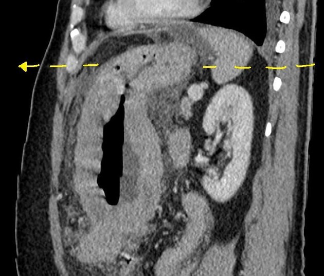
Annotated image explains the absence of the gastric air bubble as the fundal wall is extremely thickened and almost both walls oppose each other obliterating the lumen.
Case Discussion
A number of features in this case favored a diagnosis of primary gastric lymphoma, rather than secondary/systemic lymphoma or an alternative diagnosis of adenocarcinoma:
- linitis plastica appearance
- extension along the duodenum
- absence of gastric outlet obstruction symptoms
- no splenomegaly, adjacent lymphadenopathy, and normal retroperitoneum
There was also evidence of omental caking and peritoneal carcinomatosis.
Upper GI endoscopy was performed and the histopathology results yielded Burkitt lymphoma (B-cell non-Hodgkin lymphoma).
Histopathology report:
Biopsies of gastric mucosal fragments show surface ulceration and infiltration by malignant tumor cells predominantly in an interstitial pattern, composed of uniform medium-sized lymphocytes with round neuclei, finely dispersed chromatin and multiple small nucleoli. Numerous mitotic figures and apoptotic bodies are noted. Negative for H.pylori and intestinal metaplasia. Cautery artifact is noted.
Immunohistochemistry:
- The tumor cells are diffusely positive for LCA, CD20, CD79a, BCI-6 and CD10.
- Tumor cells are negative for CD3, CD5, CD23, Cyclin D1, Bcl-2, and Tdt.
- Ki-67 proliferation index is near 100%.
Diagnosis: positive involvement by high-grade Non Hodgkin's B-cell lymphoma - Burkitt type.
--
Special thanks to Dr Mohammed A. Al Sayegh.


 Unable to process the form. Check for errors and try again.