
Presentation
Upper abdominal discomfort, significant weight loss, anorexia and early satiety.
Patient Data

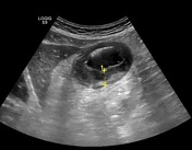

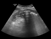
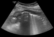
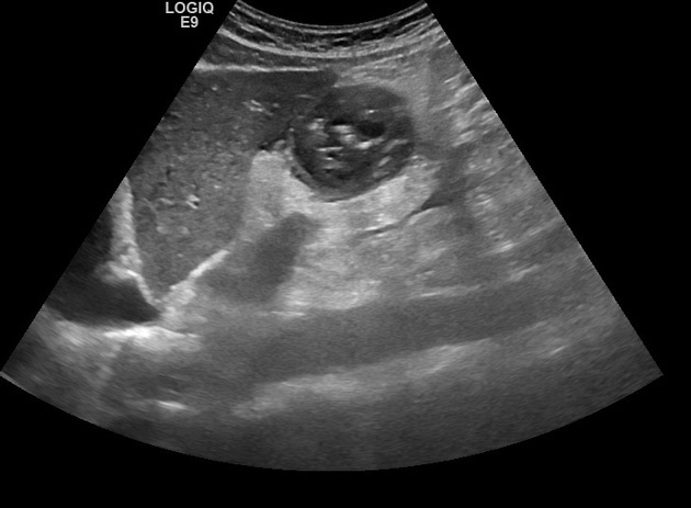
Ultrasound of the upper abdomen shows marked circumferential asymmetrical wall thickening and obscuration of the normal layers of the distal stomach. These changes extend to the proximal duodenum.
The visualized part of stomach shows some luminal narrowing.
No visible adjacent lymphadenopathy.
Pathology report:
GROSS
Specimen fixed labeled with patient’s name, consists of multiple gray white soft tissue.
MICROSCOPIC
Specimen is composed at large of necroinflammatory tissue "ulcer site", with
only one fragment showing viable gastric mucosa, showing infiltration of
lamina propria by sheets of large atypical lymphoid- like cells, with scattered
mitotic figures. Immunohistochemical stain including CD20, CD3, Ki67 and
CK are necessary for further sub-classification and to exclude undifferentiated carcinoma.
DIAGNOSIS: Stomach, biopsy: Malignant tumor, with features of high-grade non-Hodgkin lymphoma, for immunohistochemical stains.
Case Discussion
In this case, we advised further evaluation with contrast-enhanced CT, but the patient's treating physician opted for biopsy.
The stomach is the most common site of gastrointestinal tract lymphoma, extranodal lymphoma, and MALT lymphoma.
Features that can help differentiate lymphoma from gastric carcinoma is that the wall thickening is more marked in lymphoma and extension to the duodenum is typical for lymphoma. Infiltration of adjacent organs is more likely in carcinoma. However, there is an overlap in the imaging appearance of the two conditions.


 Unable to process the form. Check for errors and try again.