
Presentation
Altered mental state.
Patient Data
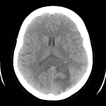
There is a left temporo-parieto-occipital lesion that transgresses the splenium of the corpus callosum. Associated vasogenic edema +/- cytotoxic edema involving the posterior parietal cortex. The most probable diagnosis is glioblastoma multiforme. There could be an associated stroke-like phenomenon, but it is most probably secondary to the tumor. MRI is suggested to confirm the diagnosis is this examination was realized without IV contrast.
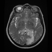

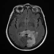
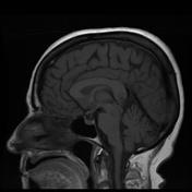
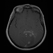
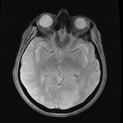

MRI confirms a large, heterogeneous, mass involving the left temporoparietooccipital lobes which extends across the splenium of the corpus callosum. There is a slight extension of the mass into the right occipital parenchyma. The lesion is partly solid, with necrotic & hemorrhagic components. Associated local mass effect & edema. The findings are compatible with glioblastoma.
Case Discussion
The diagnosis of glioblastoma was confirmed on histology. Other differential diagnoses are less plausible. Glioblastoma is the most frequent corpus callosal tumor. Another tumor that can involve the corpus callosum is lymphoma, but it is not hemorrhagic/necrotic-like this, and it is hyperdense on CT.
Note: IDH mutation status is not provided in this case and according to the 5th Edition (2021) of the WHO classification of CNS tumors, this tumor would, therefore, be designated as a glioblastoma NOS.


 Unable to process the form. Check for errors and try again.