
Presentation
Right upper quadrant pain
Patient Data

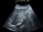
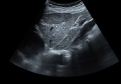
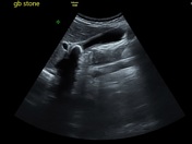
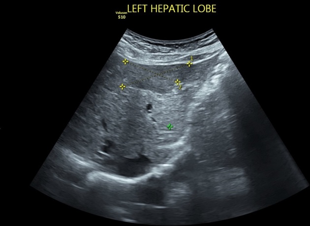
The ultrasound study revealed an incidental left hepatic lobe subcapsular relatively hypoechogenic mass. Gallbladder stone is noted presumed to be the cause of patient's complain
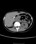

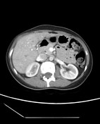
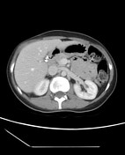
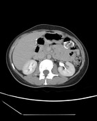

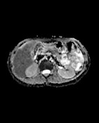
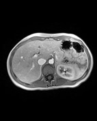
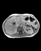
CT study revealed a left hepatic lobe well-defined ovoid lesion elicits low attenuation in the pre-contrast study. It elicits early enhancement in the late arterial phase with rapid contrast washout in the Porto-venous and delayed phases. Incidental post-cholecystectomy surgical clips

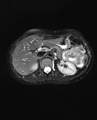
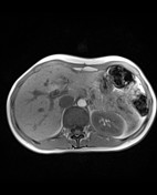
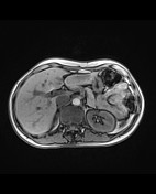
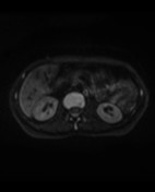
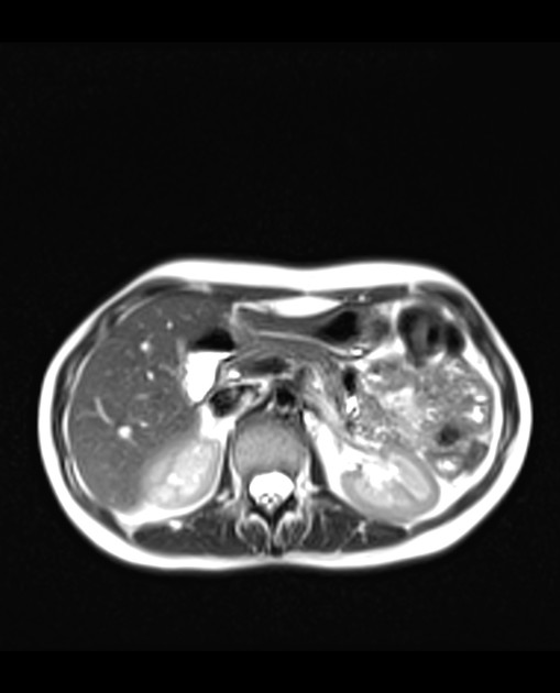
In the MRI study, the lesion is hyperintense in T2WI relative to normal hepatic parenchyma and suppressed in T2 fat sat. In T1 "in phase", the lesion is isointense to hepatic parenchyma with a significant signal drop in T1 "out of phase" denoting intracellular lipid. It elicits early enhancement in the arterial phase with contrast washout in the portal phase
Case Discussion
Here is a case of fat-containing hepatic adenoma in a middle-aged female patient receiving contraceptive pills. The enhancement pattern is similar to other hypervascular lesions (HCC, FNH, metastases), however, the incidental finding, presence of intra lesion fat, and history of pills intake, all favor the possibility of adenoma. The patient subjected to a follow up with a stationary course of this lesion developed no increased size or spontaneous rupture
Ultrasound contribution by Dr. Shaimaa Hussien


 Unable to process the form. Check for errors and try again.