Presentation
Known history of non-small cell carcinoma, evaluate for metastasis.
Patient Data
Note: This case has been tagged as "legacy" as it no longer meets image preparation and/or other case publication guidelines.
![](https://prod-images-static.radiopaedia.org/images/5735254/61e551ef0de8b8af0ee46dce8ee1fc_thumb.jpeg)
![](https://prod-images-static.radiopaedia.org/images/5735270/bf1b8293f705a7ff6ffe623154f77b_thumb.jpeg)
![](https://prod-images-static.radiopaedia.org/images/5735262/b80f21191cbfd895c80acbccd762d4_thumb.jpeg)
![](https://prod-images-static.radiopaedia.org/images/5735326/375c4846fc9e77f38e830d3072f07b_thumb.jpeg)
![](https://prod-images-static.radiopaedia.org/images/5735254/61e551ef0de8b8af0ee46dce8ee1fc_big_gallery.jpeg)
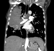
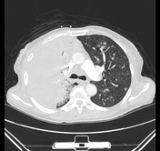
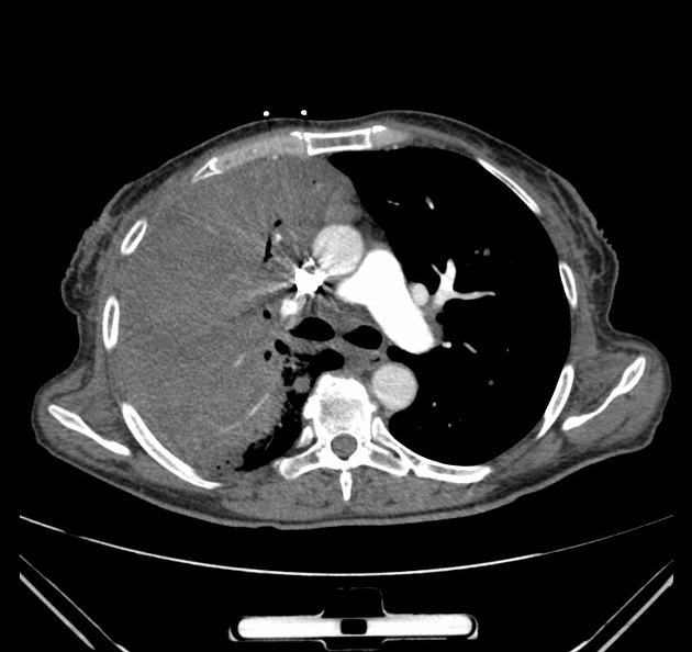
A large right lung mass with multiple smaller left lung metastases.
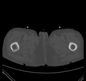
Laminated periostitis of the bilateral femora.
![](https://prod-images-static.radiopaedia.org/images/5735286/e44bc1ab2f0bb2c404d6f0dcd958de_thumb.jpeg)
![](https://prod-images-static.radiopaedia.org/images/5735294/111b7fc9040928edf9544ec6f04ea9_thumb.jpeg)
![](https://prod-images-static.radiopaedia.org/images/5735302/4465de5ac1e4f0da3e28d850a35863_thumb.jpeg)
![](https://prod-images-static.radiopaedia.org/images/5735310/00b2bbe37305a536dc10576cc956b1_thumb.jpeg)
![](https://prod-images-static.radiopaedia.org/images/5735318/754c80a21264ec3ad68bf269f97e63_thumb.jpeg)
![](https://prod-images-static.radiopaedia.org/images/5735278/ef29724d5b9d77acd84d38e077f0c3_thumb.jpeg)
![](https://prod-images-static.radiopaedia.org/images/5735286/e44bc1ab2f0bb2c404d6f0dcd958de_big_gallery.jpeg)
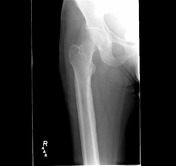
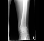
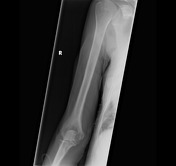
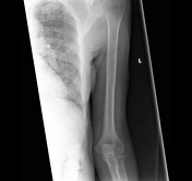
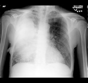
Skeletal survey demonstrates laminated periostitis of the long bones of the appendicular skeleton bilaterally. There is a large right lung opacity on the frontal chest radiograph, consistent with the patient's known non-small cell lung carcinoma.
![](https://prod-images-static.radiopaedia.org/images/5735334/dd0c4f0d0d48b79f8c95ab179859fe_big_gallery.jpeg)
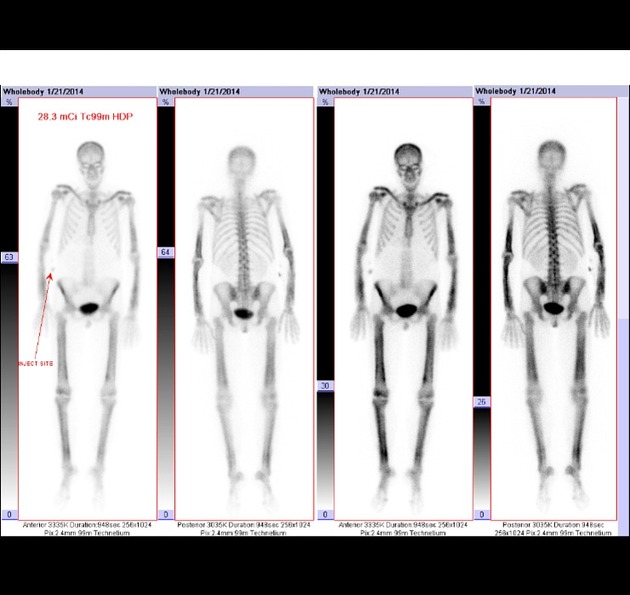
Whole body bone scan demonstrates symmetric increase uptake in the diaphyses and metaphyses of the tubular bones of the appendicular skeleton along the cortical margin, matching the findings on X-ray and CT.
Case Discussion
Patient with known non-small cell lung cancer with complaints of joint pain. Skeletal survey and whole body bone scan were performed for evaluation for metastatic disease.
Laminated periostitis of the diaphyses and metaphyses of the tubular bones of the appendicular skeleton was demonstrated.