Presentation
The patient presented with headaches for 2 months.
Patient Data
Age: 60 years
Gender: Male
From the case:
Intracranial lipoma






Download
Info
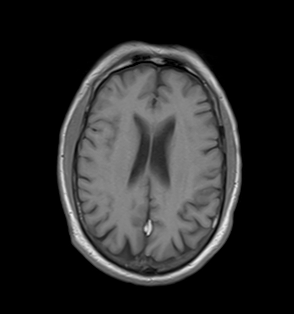
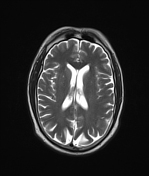
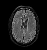
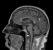
T1W hyperintense signals are seen along falx cerebri with suppression of signals on FLAIR.
From the case:
Intracranial lipoma


Download
Info
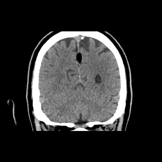
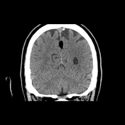
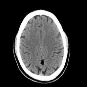
Non-contrast CT scan shows a well-defined fat density area along the falx cerebri posteriorly.
Case Discussion
Intracranial lipoma is a benign and very rare entity and is found incidentally on MRI or CT scans. Though they can be found anywhere intracranially majority are found along the midline.
They are usually asymptomatic. When symptoms do occur, they are frequently the result of co-existing general clinical conditions.


 Unable to process the form. Check for errors and try again.