
Presentation
Pre-operative chest radiograph for cholecystectomy. CT scan of chest undertaken.
Patient Data
Note: This case has been tagged as "legacy" as it no longer meets image preparation and/or other case publication guidelines.
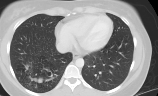
Abnormal lucencies in the right lower lobe, suggesting air trapping.
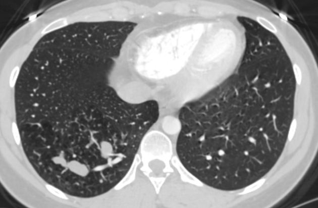
There are areas of air trapping in the right lower lobe as these areas do not communicate with the more central bronchi. Also there is increased density in the bronchiectatic bronchi, compatible with mucous plugging.
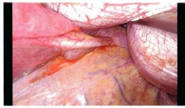
Intraoperative photograph is showing abnormal vascular supply arising from the abdominal aorta to the right lower lobe.
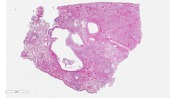
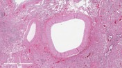

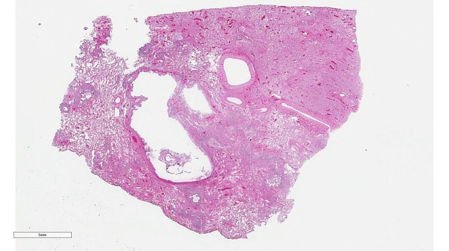
The histopathologic findings include chronic inflammatory changes including bronchiectasis, dilatation of bronchioles, foci of necrosis, acute and chronic inflammatory infiltrates, and abnormally thick-walled blood vessel(s), also consistent with intralobar sequestration.
Slides provided by Paul W. Biddinger, MD
Professor of Pathology
Case Discussion
Patient underwent right lower lobectomy without complications.
Case courtesy of Dr.William Bates.


 Unable to process the form. Check for errors and try again.