Presentation
Toddler who was generally unwell.
Patient Data


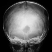
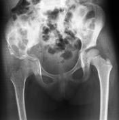
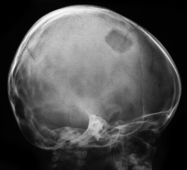
Lucent lesions in the skull and right ilium. L3 vertebra plana.
SKULL LESION BIOPSY
GROSS
The specimen is received in formalin, labeled with patient data and designated "lt parietal skull lesion". It consists of a few pieces of hemorrhagic tissue 1.5 x 1 x 0.4 cm in aggregate.
MICROSOPY
The specimen comprises sheets of histiocytic cells admixed with eosinophils, neutrophils, plasma cells, and multinucleated giant cells. The histiocytic cells frequently have indented nuclei, single nucleoli, and abundant pale cytoplasm. Necrosis and hemorrhage are also present.
Immunohistochemistry shows the histiocytic cells positively stains with CD1a and S100.
DIAGNOSIS
LEFT PARIETAL BONE OF SKULL, CURETTAGE: LANGERHANS CELL HISTIOCYTOSIS.
Case Discussion
Multiple bone lesions in keeping with Langerhans cell histiocytosis. The skull lesions show the classical bevelled edges. A single skull lesion would be termed eosinophilic granuloma.
Vertebra plana is another feature, although a differential for this in isolation exists.


 Unable to process the form. Check for errors and try again.