
Presentation
Abdominal pain for the past 3 days. Outside evaluation revealed doubtful renal calculi and was given symptomatic treatment. Since pain was not subsiding, she was referred to a higher center. Bilious vomiting was observed at the time of sonographic study of abdomen.
Patient Data

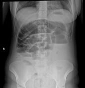

Dilated small bowel loops with multiple air-fluid levels. Dilated colon with tapering in the left abdomen.
Ultrasound correlation revealed dilated small bowel loops and large bowel up to the distal descending colon. Further evaluation with CT was considered warranted.


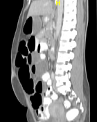
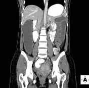
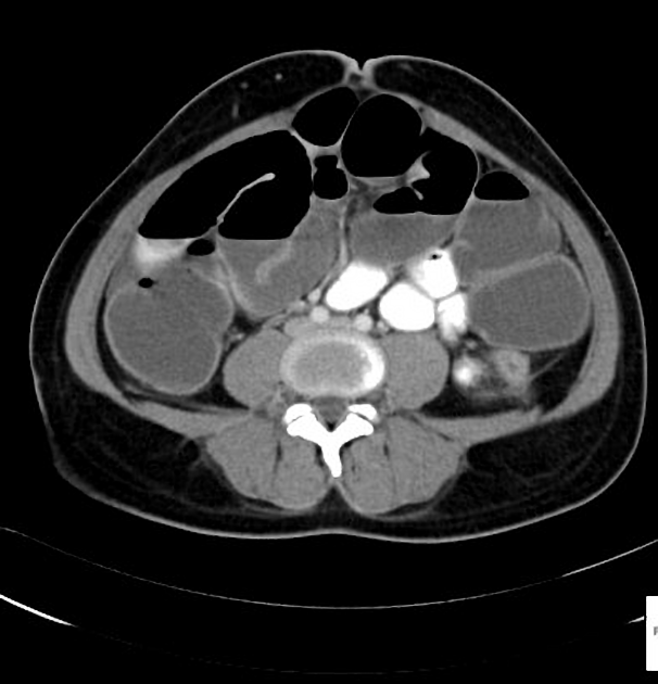
A short segment narrowing measuring 3 cm in the distal descending colon with upstream colonic dilatation with mildly enhancing wall. Dilated small bowel loops with air-fluid levels also present.
Case Discussion
A colonoscopy revealed a lumen occluding circumferential proliferative growth in the descending colon, and a biopsy was taken.
Histopathology
Microscopic Description: Section shows fragments of large intestinal mucosa containing part of an ulcerating neoplasm with villous architecture in places lined by cells displaying nuclear crowding, stratification and hyperchromasia, variably conspicuous nucleoli and frequent mitosis. Disorganized glands with luminal necrosis and moderate to severe degree of cytological atypia and apical mitosis are present in the lamina propria and also between muscularis mucosal fibers. There is associated inflammation and desmoplasia
Conclusion: Moderately differentiated colon adenocarcinoma.


 Unable to process the form. Check for errors and try again.