
Presentation
Two days history of upper abdominal pain.
Patient Data
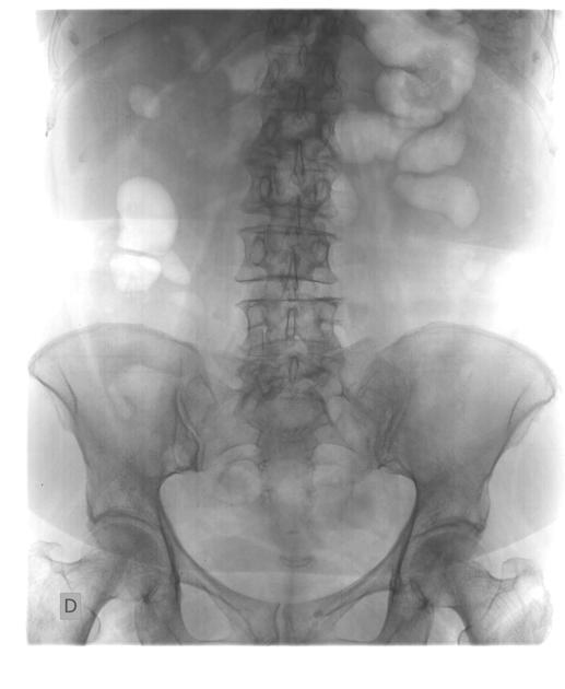
Frontal abdominal x-ray shows no abnormalities.


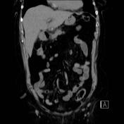
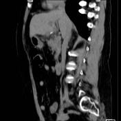
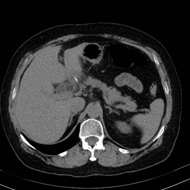
CT shows a hyperattenuating linear foreign body that punctures the lowers segments of hepatic left lobe; an hypoattenuating subcapsular small area is identified around it on liver. There is also fat planes densification between the duodenum and the liver. No pneumoperitoneum or gas on retroperitoneum.
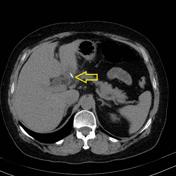
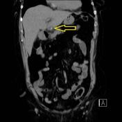
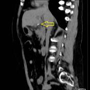
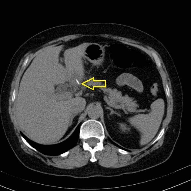
CT shows a hyperattenuating linear foreign body that punctures the lowers segments of hepatic left lobe; an hypoattenuating subcapsular small area is identified around it on liver.
Case Discussion
The case was reported as a foreign body that probably perforated duodenum and punctured left hepatic lobe causing a small liver abscess around it. The case was confirmed by surgery, the foreign body was a fishbone.


 Unable to process the form. Check for errors and try again.