
Lung abscess with subcutaneous emphysema, pneumothorax and pneumopericardium
Presentation
Patient presented with cough, fever and chest pain.
Patient Data
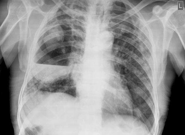
Large cavitating lesion with air-fluid level in the right mid-zone, suggestive of a lung abscess. Air space opacities and small cavities are present in both lungs.
The right costophrenic angle is not blunt due to effusion (confirmed on ultrasonography, not shown here), but due to consolidation and fibrotic changes.
Mild elevation of the right hemidiaphragm.
Post intercostal drainage tube

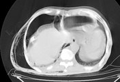

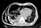
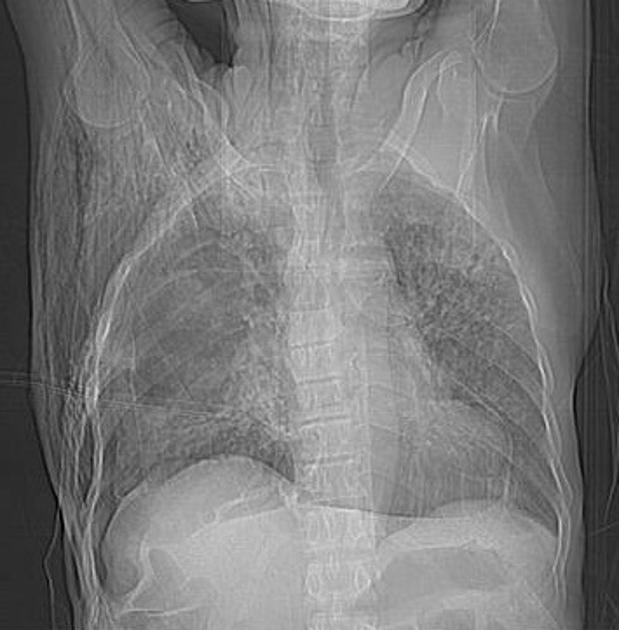
Surgical emphysema, pneumothorax and pneumopericardium (probably due to pleuro-pericardial fistula which is a complication of infection) developed after ICD insertion.
Large right lung abscess (with ICD tube in-situ) and multiple cavities in both lungs.
Case Discussion
A diagnosis of lung abscess was made on chest x-ray. A large bore intercostal drainage tube was inserted. During the procedure the tube migrated a little which was then re-positioned. Following this there was visible subcutaneous air accumulation and the patient became tachypneic. The patient was stabilized and CT scan was performed which showed surgical emphysema, pneumothorax and pneumopericardium.


 Unable to process the form. Check for errors and try again.