Presentation
Right hip pain and back pain
Patient Data
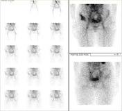
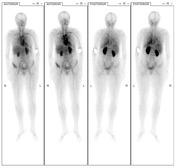

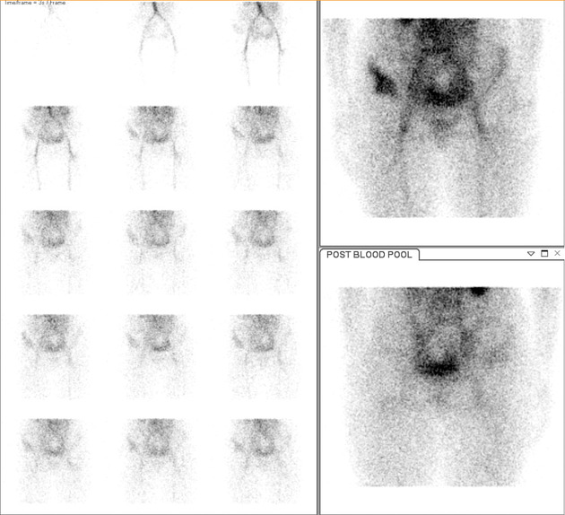
On early phase blood flow and blood pool imaging, there is hyperemia in the region of the right anterior iliac bone.
On delayed phase planar imaging, there is heterogeneous increased tracer accumulation in the right anterior iliac bone superior to the acetabulum. Inferior to the increased uptake, there is a photopaenic region. Also, there is mildly increased tracer accumulation in the pedicles of the T12 vertebral body, best seen on the posterior views.
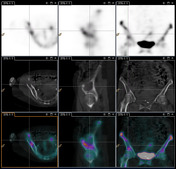
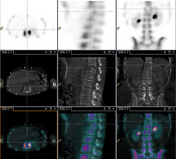
SPECT/CT of the pelvis demonstrates a lytic lesion in the right iliac bone, which photopaenia in the region of the lesion and increased tracer uptake at the periphery. In addition, there is a second lytic lesion in the anterior T12 vertebral body which is cold on bone scan.
Case Discussion
This patient has a history of breast cancer with lytic metastases of the right pelvis and T12 vertebral body. As tracer uptake on bone scan reflects osteoblastic activity and matrix deposition, lytic metastases are often photopaenic. Increased tracer accumulation can be seen at the edge of the lesion reflecting the reactive changes in the bone adjacent the destructive lesion, as seen in this case. Breast cancer metastases can demonstrate variable uptake on bone scan.


 Unable to process the form. Check for errors and try again.