
Presentation
Evaluation of incidental abdominal mass.
Patient Data
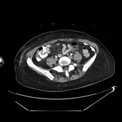

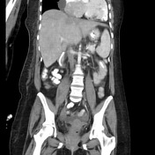
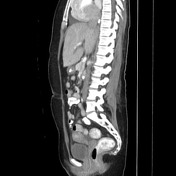

Small oval central mid mesenteric mass with well-circumscribed borders.
Cardiomegaly with small pericardial effusion. Feeding tube. Multifocal R>L renal cortical scaring. Small ascites.
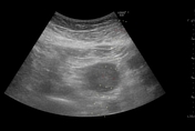
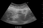
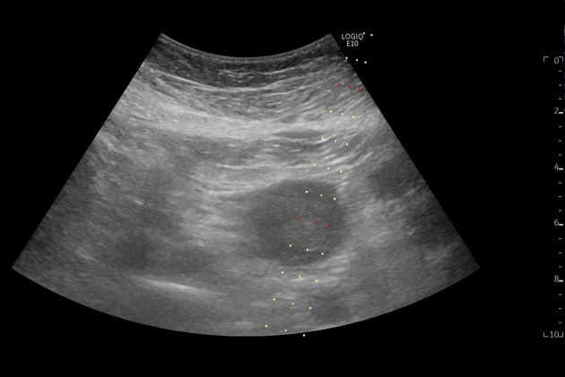
Rounded mesenteric mass approximated to the body wall with manual pressure, displacing the overlying small bowel loops. Center of mass measures 10 cm with the probe guide in place. Mass successfully biopsied.
Case Discussion
The small mass underwent ultrasound-guided biopsy which confirmed a diagnosis of demoed-type fibromatosis.
It might surprise you that this can be safely biopsied with ultrasound given the relatively deep position in the mesentery. However, the small bowel loops can often be displaced away from the mass/body wall with gentle compression, essentially bringing the mass up to the body wall. Careful inspection for any peristalsing loops of bowel can be made before advancing the biopsy device. In you are highly concerned by this, a biopsy device can safely pass through small bowel if needed (20 gauge or smaller is recommenced; note that traversing large bowel is higher risk and prep and antibiotics may be considered 1). Thus, ultrasound is actually much safer for this type of biopsy than CT, which would not be safe in this case.


 Unable to process the form. Check for errors and try again.