
Presentation
ED presentation with three day history of abdominal pain and distension. ? bowel obstruction.
Patient Data


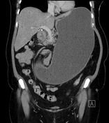
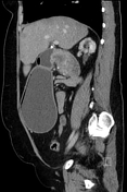
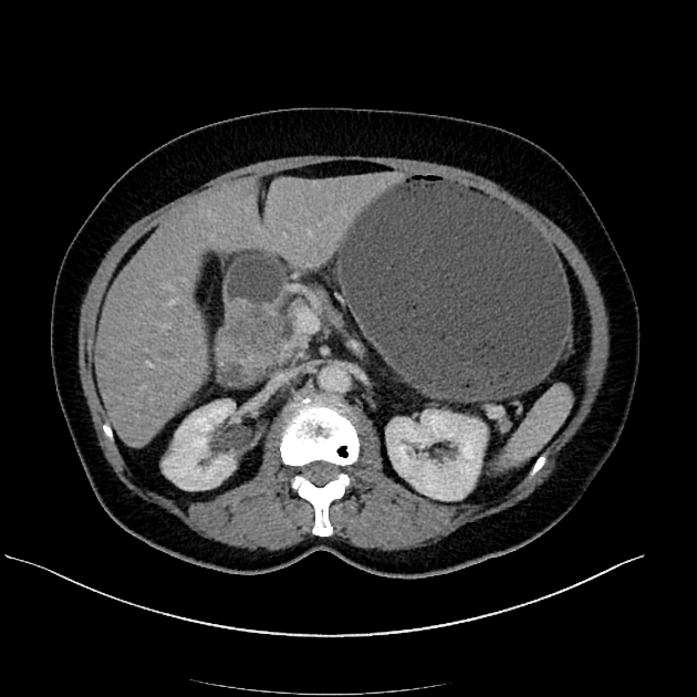
The stomach is massively distended and fluid-filled. There is abrupt luminal narrowing within D2 where there is a circumferential soft tissue density lesion seen. Small foci of internal calcification are seen. The bowel is collapsed distal to this site.
Low-density, ill-defined liver lesions are evident. The largest lesion is located within segment IVa.
An enlarged porta hepatis node is seen.
Overall appearance is highly suspicious for a primary duodenal malignancy with local lymphadenopathy, hepatic metastases and duodenal obstruction. Pancreatic head or ampullary malignancy is considered less likely given the absence of biliary or pancreatic duct dilatation.
Case Discussion
The patient underwent upper GI endoscopy and biopsy which was consistent with duodenal adenocarcinoma.
Adenocarcinoma is the most common malignancy of the duodenum. Half of patients will have metastatic disease at the time of presentation.


 Unable to process the form. Check for errors and try again.