
Presentation
Abdominopelvic pain and chronic constipation. Bright red blood in the stool. Weight loss
Patient Data


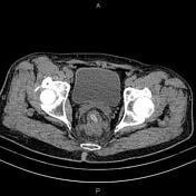

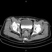
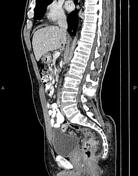
Asymmetrical increased wall thickness due to tumoral infiltration is present at the rectum. Mesorectal fascia is infiltrated at the right posterior aspect. Perirectal fat stranding and several enlarged lymph nodes with SAD less than 12 mm are also evident.
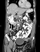
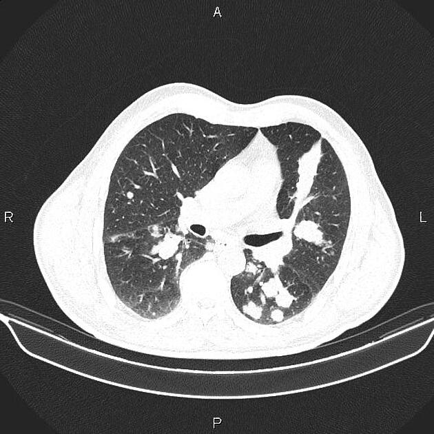
Multiple nodules are seen at both lung fields less than 30 mm, consistent with metastases. In addition, an atelectatic band is evident in the left lingula segments.
A few enlarged lymph nodes are seen in both hilar regions.
The right hemidiaphragm is elevated.
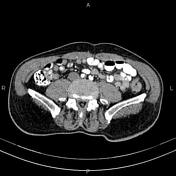
At least three small low-enhancing masses are seen at the liver less than 18 mm, suspected for metastases. Additionally, extra and central intrahepatic bile ducts are dilated.
The prostate gland is enlarged.
Case Discussion
Rectal mass, pathology-proven adenocarcinoma with mesorectal fascia invasion, regional lymphadenopathy, multiple pulmonary and a few hepatic metastases.


 Unable to process the form. Check for errors and try again.