
Abnormal rectal cancer nodes
These morphologic criteria assess whether locoregional lymph nodes may be involved in rectal cancer.
Original work of Niharika Praveen.
Case Discussion
In rectal cancer, high resolution MRI can be used for staging.
For tumors above the dentate line, the short axis of nodes in the mesorectal, superior rectal and inferior mesenteric territories is measured.
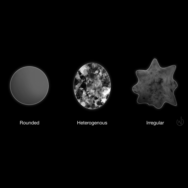
Three morphologic criteria are also assessed 1:
heterogeneity/irregularity of signal
irregularity of border
round shape
The determination of whether nodes are involved by disease or not is made as follows:
>9mm in short axis diameter = suspicious
5-9mm in short axis diameter and meet 2 out of 3 criteria = suspicious
<5mm and meet all 3 criteria = suspicious
Following chemoradiotherapy, nodes measuring greater than 5 mm in short axis diameter are considered to be suspicious.
Note that these criteria do not apply to internal iliac or obturator nodes, which are considered as locoregional but have different size thresholds. They also do not apply to external iliac, common iliac. paraaortic and inguinal nodes (for tumors above the dentate line); these are considered metastatic (M).
Case uploaded with assistance from Dr Vikas Shah.


 Unable to process the form. Check for errors and try again.