
Presentation
Rule out pneumonia. Patient is previously known for left upper lobe adenocarcinoma.
Patient Data

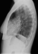
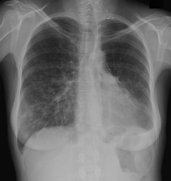
Chest x-ray shows bilateral, but mainly right-sided small airspace opacities. Some of the opacities are cavitary. Left pleural effusion. Left upper lobe resection is seen.
A metastatic process must be excluded, since the patient has already had lung cancer. An infectious or inflammatory process is also possible. Chest CT-scan is recommended.
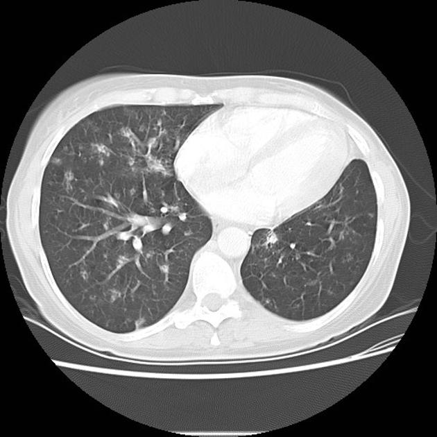
Chest CT-scan confirms multiple small bilateral cavitary opacities surrounded by ground glass. The findings are not specific; an infectious process can be considered, but it is atypical. Multifocal adenocarcinoma must also be considered. Metastases are also possible.
Case Discussion
The histological diagnosis of adenocarcinoma was later confirmed, in this patient previously known for left upper lobe adenocarcinoma.


 Unable to process the form. Check for errors and try again.