
Presentation
Palpable non painful left neck swelling
Patient Data
Age: 40 years
Gender: Male
From the case:
Neck intermuscular lipoma


Download
Info
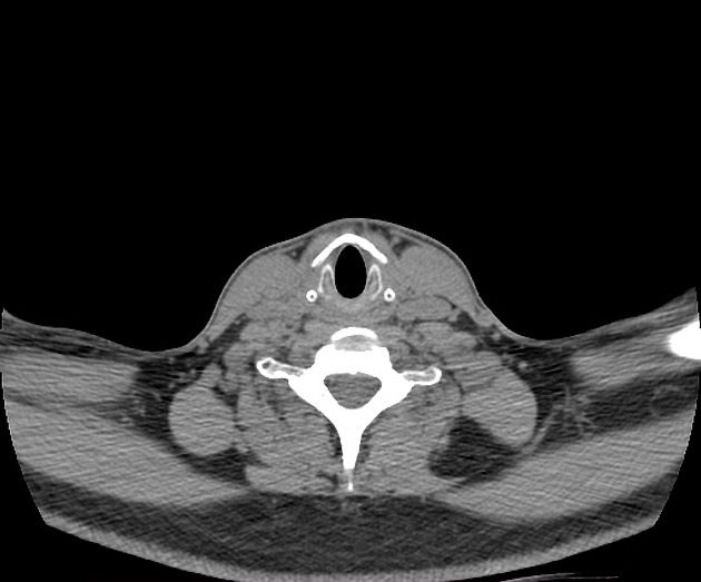
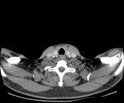
An elliptical intermuscular homogeneous hypodense mass with few thin non-enhancing internal septa seen at the left lower posterolateral aspect of the neck between the trapezius muscle posteriorly, levator scapulae muscle anterolaterally and deep cervical (paraspinal) muscles medially. It measures about 2.5 x 5 x 5 cm in AP x TR x CC respectively and about -96 HU in density.
Case Discussion
CT features are most consistent with an intermuscular lipoma.
The most common differential diagnosis is well-differentiated liposarcoma which is usually bigger in size (>10 cm) and usually shows percentage fat <75%, thick or nodular (>2 mm) septa, nodular/globular areas and prominent enhancement.


 Unable to process the form. Check for errors and try again.