
Presentation
Newborn with antenatally detected left renal abnormality.
Patient Data

Three discrete solid appearing foci are seen within the left kidney. They are homogeneously echogenic with the largest within the mid point measuring 27 x 23 x 29 mm. These are vascular in nature. No hydronephrosis.
The right kidney and bladder (not shown) were normal.


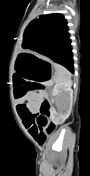
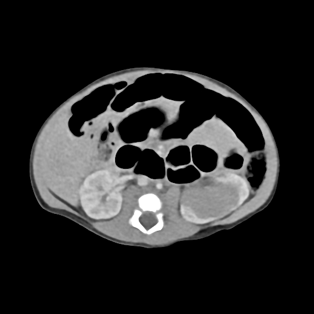
Three hypodense left renal masses with minimal post contrast enhancement.
No right sided lesion identified. No nodal disease or distant metastases.
Left ureteronephrectomy was performed.
HISTOLOGY
Macroscopic:
There are multiple lesions and smaller nodules. All lesions are similar, with a cream granular surface with no evidence of necrosis or hemorrhage. There is no involvement of the vascular margin or of the ureter.
Microscopic:
The sections of kidney show multinodular nephroblastomatosis. There are multiple perilobar nephrogenic rests throughout the kidney, many of which are microscopic in size. The macroscopically visible nodules represent hyperplastic nephrogenic rests. All nodules show a sharp demarcation with the adjacent kidney and there is no compression of adjacent structures, with native glomeruli present and just intermingling with the periphery of the nodules. None of the nodules have a fibrous capsule.
Summary:
Nephroblastomatosis
Case Discussion
Nephroblastomatosis represents persistence of the primitive metanephric blastema.
There are two pathologic subtypes of nephrogenic rest: perilobar and intralobar.
There is the potential for malignant transformation to Wilm's tumor - mandating close follow-up or treatment.


 Unable to process the form. Check for errors and try again.