Neuroanatomy - lateral cortex (illustrations)

Case contributed by Frank Gaillard
Diagnosis not applicable

Lobes of the brain

Illustration
This study is a stack
Lobes
This study is a stack
Lobes &
insula
This study is a stack
Frontal
lobe
This study is a stack
Parietal
lobe
This study is a stack
Temporal
lobe
This study is a stack
Occipital
lobe
Download
Info

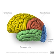
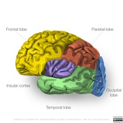
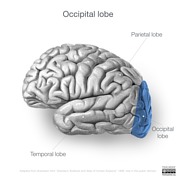
Images of the lobes of the brain, the insula cortex (not considered a true lobe by most, but included here for completeness), and their lobar and sulcal relationships. 

Note: multiple versions of each image are provided as a stack. 

Sulci of the brain and lobes

Illustration
This study is a stack
Brain
This study is a stack
Frontal
lobe
This study is a stack
Frontal
lobe
This study is a stack
Parietal
lobe
This study is a stack
Parietal
lobe
This study is a stack
Temporal
lobe
This study is a stack
Temporal
lobe
This study is a stack
Occipital
lobe
This study is a stack
Occipital
lobe
Download
Info

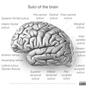
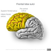
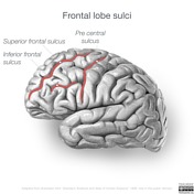
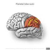
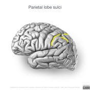
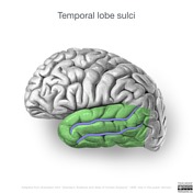
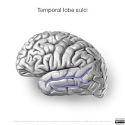
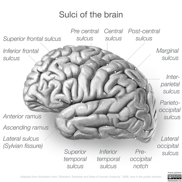
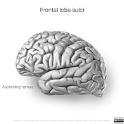
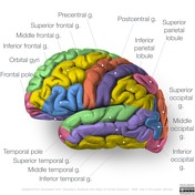
Images depicting the sulci of the whole brain and for each lobe. 

Note: multiple versions of each image are provided as a stack. 

Individual sulci

Illustration
This study is a stack
Superior
frontal sulcus
This study is a stack
Inferior
frontal sulcus
This study is a stack
Anterior and
ascending rami
This study is a stack
Precentral
sulcus
This study is a stack
Central
sulcus
This study is a stack
Post-central
sulcus
This study is a stack
Interparietal
sulcus
This study is a stack
Marginal
sulcus
This study is a stack
Parieto-occipital
sulcus
This study is a stack
Lateral
sulcus
This study is a stack
Superior
temporal sulcus
This study is a stack
Inferior
temporal sulcus
Download
Info

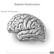
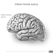
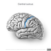
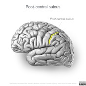
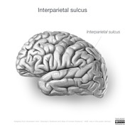
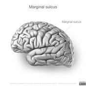
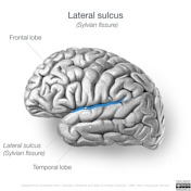
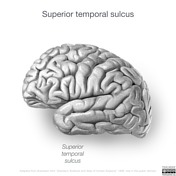
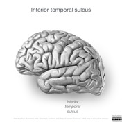
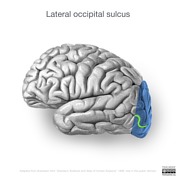
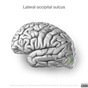
Images depicting each individual sulcus. 

Note: multiple versions of each image are provided as a stack. 

Gyri and lobules

Illustration
This study is a stack
Gyri
This study is a stack
Frontal
lobe gyri
This study is a stack
Frontal
pole
This study is a stack
Superior
frontal gyrus
This study is a stack
Inferior
frontal gyrus
This study is a stack
Middle
frontal gyrus
This study is a stack
Frontal
operculum
This study is a stack
Precentral
gyrus
This study is a stack
Orbital
gyri
This study is a stack
Pre and
Postcentral gyri
This study is a stack
Postcentral
gyrus
This study is a stack
Pareital
lobe gyri
This study is a stack
Superior
partietal lobule
This study is a stack
Inferior
parietal lobule
This study is a stack
Angular
gyrus
This study is a stack
Supramarginal
gyrus
This study is a stack
Temporal
lobe gyri
This study is a stack
Temporal
pole
This study is a stack
Middle
temporal gyrus
This study is a stack
Superior
temporal gyrus
This study is a stack
Inferior
temporal gyrus
This study is a stack
Occipital
lobe gyri
This study is a stack
Superior
occipital gyrus
This study is a stack
Middle
occipital gyrus
This study is a stack
Inferior
occipital gyrus
Download
Info

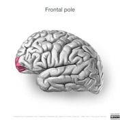
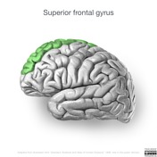
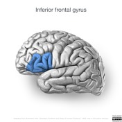
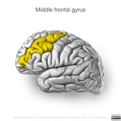
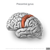
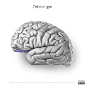
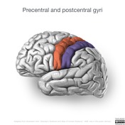
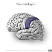
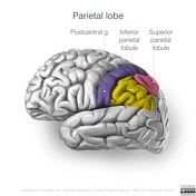
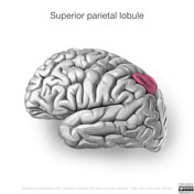
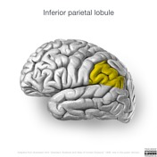
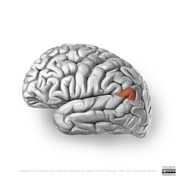
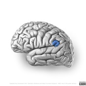
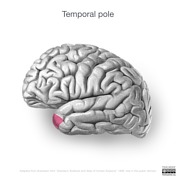
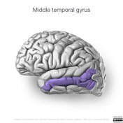
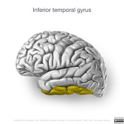
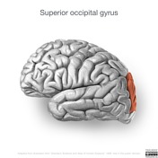
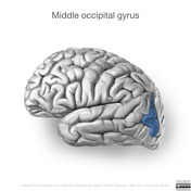
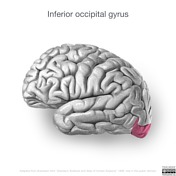
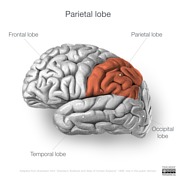
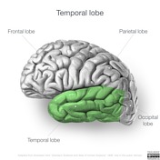
Images depicting individual regions. 

Note: multiple versions of each image are provided as a stack. 

Case Discussion

These illustrations are based on the diagrams from "Sobotta's Textbook and Atlas of Human Anatomy 1908", the copyright of which has lapsed. The source files were obtained from wikimedia commons, and are listed below. 

The images have been edited to remove labels, change contrast and slightly tweak some details. In some instances more than one image has been combined (for example the cut-away views of the insular cortex). 

Source files

  1. insular cortex: https://commons.wikimedia.org/wiki/File:Sobo_1909_633.png
  2. lateral brain: https://commons.wikimedia.org/wiki/File:Sobo_1909_626.png

How to use cases

You can use Radiopaedia cases in a variety of ways to help you learn and teach.

Creating your own cases is easy.

:

Updating… Please wait.

 Unable to process the form. Check for errors and try again.

 Thank you for updating your details.