
Presentation
Pain post eating.
Patient Data


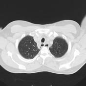
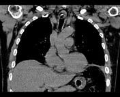
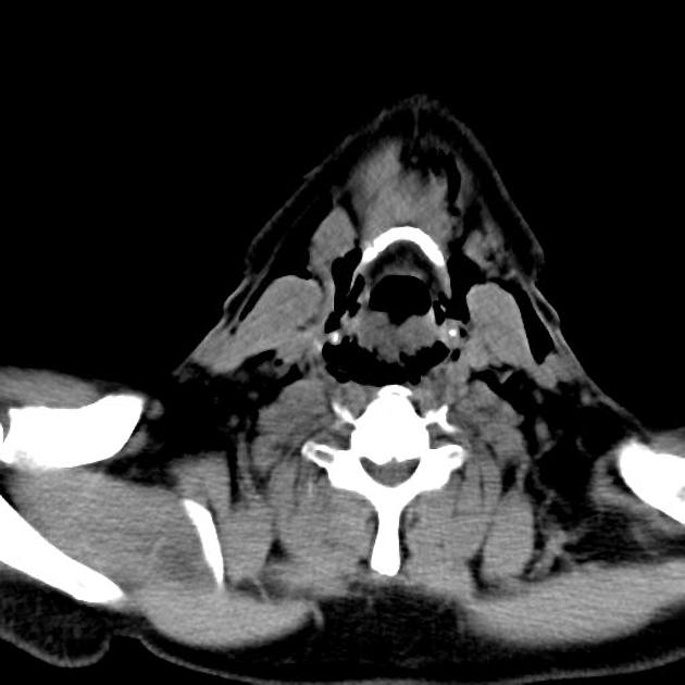
Pneumomediastinum, with paraesophageal/posterior mediastinal gas tracking superiorly around the infrahyoid strap muscles. Small bilateral pleural effusions, marginally larger on the left, with associated medial bibasal collapse.
An obliquely oriented, 2.1cm long X 0.4cm wide, curvilinear hyperdensity previously seen within the superior mediastinal esophagus, anteriorly adjacent to T1/2.
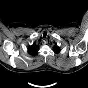
Within the imaged upper abdomen, a small fluid-density collection is seen around to gallbladder and hepatorenal pouch, however, there is no evidence of gallbladder wall thickening or gallstone identified. A very small fluid density collection is noted within the right anterior perirenal space. Upper abdominal organs are of unremarkable appearance.
Conclusion:
- Pneumomediastinum, reduced/redistributed since the previous examination.
- 2.1cm curvilinear hyperdensity may represent a foreign body.
Case Discussion
Culprit turned out to be swallowed chop bone.


 Unable to process the form. Check for errors and try again.