
Presentation
Re-staging bone scan for advanced metastatic prostate carcinoma under hormone suppression therapy. Low back pain and headaches. PSA 250 ng/ml.
Patient Data
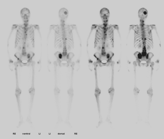
The bone scan shows (among others) a large circumscribed activity zone in the right posterior calvarium due to disseminated bone metastases.
Images courtesy of Caroline Mommsen, MD
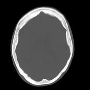

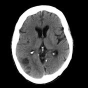


Pre- and post-contrast soft window CT images of the brain show a hypervascular mass extending from the calvarium into the brain. The borders towards the brain are irregular, suggesting direct invasion rather than displacement (Fig. 2). There are liquid inclusions at the tumor periphery, probably from previous hemorrhage.
Case Discussion
Prostate cancer, in spite of its distant location, is the second most common primary tumor metastasizing to the skull. Invasion beyond the dural border into the neuroaxis is however extremely rare.


 Unable to process the form. Check for errors and try again.