Presentation
Abdominal pain, with a clinically palpable large abdominal mass.
Patient Data
![](https://prod-images-static.radiopaedia.org/images/62841495/c0c83b5c116b445a9c2c73ff18746e48efce7b7e5ac5930b255651a1c50d5564_thumb.jpeg)
![](https://prod-images-static.radiopaedia.org/images/62841496/a3a704c878d42ff4bd4ed2dd608ace69fb533958cfe81534385d605ddc5e5ec1_thumb.jpeg)
![](https://prod-images-static.radiopaedia.org/images/62841495/c0c83b5c116b445a9c2c73ff18746e48efce7b7e5ac5930b255651a1c50d5564_big_gallery.jpeg)
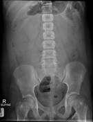
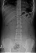
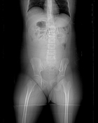
There is a paucity of bowel gas, with peripherally displaced and collapsed large bowel suggestive of a central abdominal mass. The psoas shadows are partly maintained. There is minimal fecal loading. The abdominal skeleton is normal.
Incidental clothing artefact, left groin, (an aglet).
Courtesy: Dr S Palliam.
![](https://prod-images-static.radiopaedia.org/images/62841497/cd6e7ca45194e87e558a2e511d29b61fcb6a42a72397f1472267bdaf5970aa9c_big_gallery.jpeg)
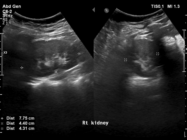
There is a large, central, abdominopelvic multicystic mass. There is no renal tract obstruction, no ascites and no hepatic metastases or lymph adenopathy.
The left ovary and uterus are normal.
The right ovary appeared normal on ultrasound.
Courtesy: Dr S Palliam
![](https://prod-images-static.radiopaedia.org/images/62841510/7b040471a3d19b67edd3a67a46b7a9a159d3b6c3edb00cc6eb6fc2fa859c73cf_thumb.jpeg)
![This study is a stack](/packs/stack-YQKLCKBI.gif)
![](https://prod-images-static.radiopaedia.org/images/62841749/7ae2842836c139ebb86e18affdc269b350cbeb3fa987154978b5085e966ae65e_thumb.jpeg)
![This study is a stack](/packs/stack-YQKLCKBI.gif)
![](https://prod-images-static.radiopaedia.org/images/62841818/716264a208be5cb38b7c54cda10bdbc9445ca5e6290ca7c59c548ec14de4f2ad_thumb.jpeg)
![This study is a stack](/packs/stack-YQKLCKBI.gif)
![](https://prod-images-static.radiopaedia.org/images/62841646/35fda2c5ee08561a44bfb26eaab50698ae0ce8395fc4f567d519d84d9761a7e3_thumb.jpeg)
![This study is a stack](/packs/stack-YQKLCKBI.gif)
![](https://prod-images-static.radiopaedia.org/images/62841561/14be0979c79f09db9155579266c585d183238b14cdfc8f6812841f056a0fdf02_thumb.jpeg)
![This study is a stack](/packs/stack-YQKLCKBI.gif)
![](https://prod-images-static.radiopaedia.org/images/62841510/7b040471a3d19b67edd3a67a46b7a9a159d3b6c3edb00cc6eb6fc2fa859c73cf_big_gallery.jpeg)
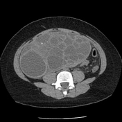
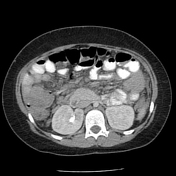
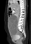
CT demonstrates a large, encapsulated, multiseptated, predominantly cystic mass lesion arising from the right adnexa. Minimal flecks of calcification are present. There is minimal enhancement of the capsule and septa with the absence of any solid or suspicious component. There is a noted absence of fat and dental elements. The uterus and left adnexa are grossly normal.
There are displaced small and large bowel loops with no bowel obstruction.
There is no renal tract obstruction.
There is free fluid within the pelvis.
There are no features to suggest metastases.
Courtesy: Dr DH JOGI.
![](https://prod-images-static.radiopaedia.org/images/62841879/08d87e16c57a270a2e55a7eae17ba34bce7ea2eac2e6585d7e294a12f3abfaa6_thumb.jpeg)
![](https://prod-images-static.radiopaedia.org/images/62841880/feeebd9af0b634a58b937d93fc6b40c25c5b69962794859604566db2c9a02395_thumb.jpeg)
![](https://prod-images-static.radiopaedia.org/images/62841879/08d87e16c57a270a2e55a7eae17ba34bce7ea2eac2e6585d7e294a12f3abfaa6_big_gallery.jpeg)
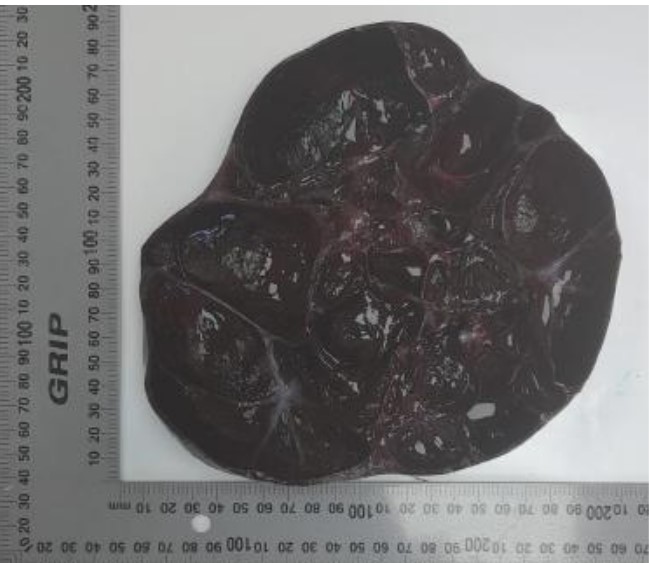
Gross specimen of the right ovarian cystectomy. The ovary/ovarian cyst measured 200 x 200 x 95 mm in dimensions. There was macroscopic evidence of torsion, with a multilocular and hemorrhagic appearance.
Histology excluded malignancy and endometriosis with complete infarction of the specimen secondary to torsion.
Case Discussion
A histopathologically proven large, multiseptated, benign, right ovarian cyst and ovarian torsion.
The broad differential diagnosis includes: