
Presentation
Left renal tumor.
Patient Data
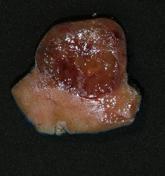
This is a papillary renal cell carcinoma removed by partial nephrectomy.
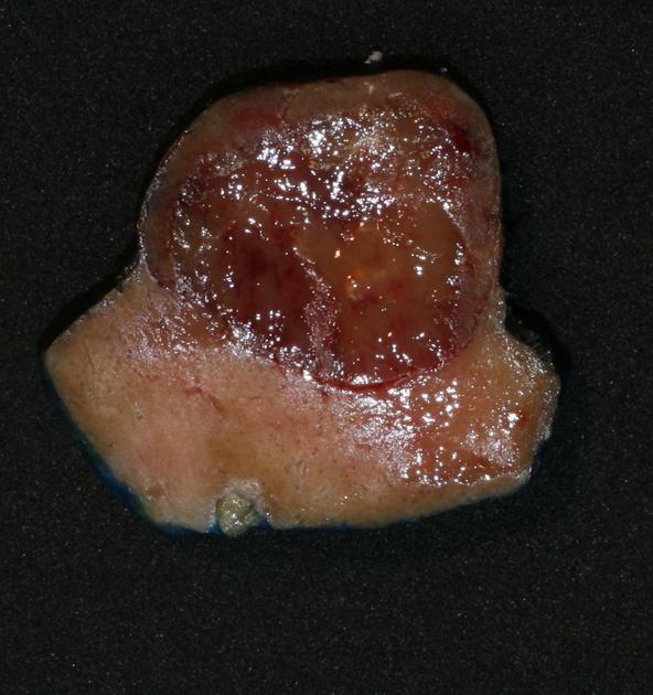
Macroscopic - Circumscribed, cortical-based, tan tumor with a cuff of renal parenchyma.
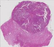
Microscopic (low power) - circumscribed lesion.
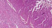
Microscopic (medium power) - well-defined interface of tumor and adjacent renal cortex (bottom left).
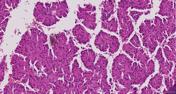
Microscopic (high power) - well-formed branching papillae with lining cells showing abundant eosinophilic cytoplasm and small uniform nuclei.
Case Discussion
The features are consistent with a papillary renal cell carcinoma.
Papillary RCC is further subclassified on morphological and prognostic grounds into type I and type II; this is a good example of a type II papillary RCC with cores lined by larger lining cells with abundant eosinophilic cytoplasm, arranged in a 'pseudostratified' pattern (in areas the cells appear to be several layers thick, but are infact all still attached to the basement membrane).


 Unable to process the form. Check for errors and try again.