
Parasternal short axis view - normal (transthoracic echocardiography)
Presentation
Asymptomatic male.
Patient Data
Parasternal Short Axis
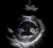

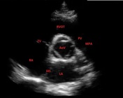

Images obtained with a low frequency phased array transducer in the third left intercostal space, with the probe marker rotated 90° clockwise from the parasternal long axis, then fanning in a cephalad direction.
Image 1 (blue labels) shows a normal parasternal short axis at the level of the papillary muscles
Image 2 (red labels) shows a normal parasternal short axis at the mitral valve level
Image 3 (red labels) shows a normal parasternal short axis at the base of the heart
Abbreviations: LA (left atrium), RA (right atrium), IAS (interatrial septum), MV (mitral valve), TV (tricuspid valve), ALMV (anterior leaflet, mitral valve), PLMV (posterior leaflet, mitral valve), LV (left ventricle), IW (inferior wall, left ventricle)IVS (interventricular septum), PWLV (posterior wall, left ventricle), PmP (posteromedial papillary muscle), AIP (anterolateral papillary muscle), AoV (aortic valve), RVOT (right ventricular outflow tract), BPW (basal posterior wall, left ventricle), PCD (pericardium), PV (pulmonary valve), MPA (main pulmonary artery = pulmonary trunk)
Case Discussion
Normal scan for reference.


 Unable to process the form. Check for errors and try again.