
Presentation
Fall from stairs
Patient Data


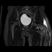
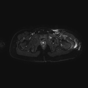
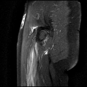
A well-defined rounded intramedullary STIR hyperintense lesion in the proximal femur shaft was noted, suggesting a mass lesion.
Complete, displaced left subtrochanteric femoral fracture at the level of the lesion.
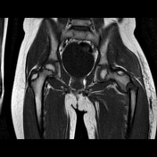
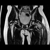
The left gluteus medius appears bulky with a nearly complete tear in the distal portion with retraction of muscle fibers.
Edematous changes in the insertional portion of the left gluteus minimus, with intact fibers.
Diffuse edema in the adjacent soft tissue.
STIR hyperintense marrow signal in the proximal femoral shaft.
Case Discussion
The MRI findings suggesting intramedullary lesion with associated pathological subtrochanteric femur fracture. No follow-up is available as the patient was referred to another hospital.
Pathological fractures are typically reserved for fractures through abnormal bone due to tumors. The most typical location for pathological fractures is the subtrochanteric region of femur ref.


 Unable to process the form. Check for errors and try again.