
Presentation
Increasing abdominal distension and discomfort. Some nausea.
Patient Data
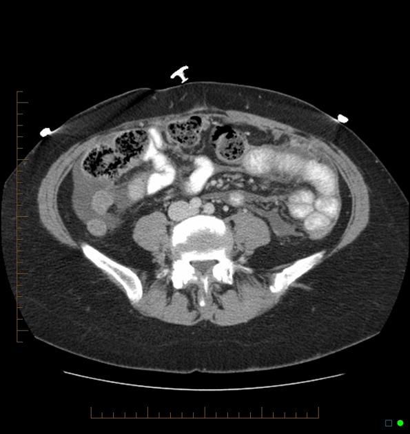
Extensive fluid is seen in the peritoneal cavity associated with thickening of the peritoneum which enhances and appears nodular in places. The omentum, located anteriorly and to the left of the midline, appears stranded and bulky with multiple nodular regions of soft tissue density.
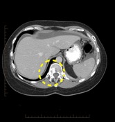
Nodular pleural thickening is noted on the left, and bilateral breast prostheses in situ. On the right side of the T12 vertebral body, involving the posterior vertebral body and right pedicle, is a region of mixed sclerosis and lysis. Similar other regions are present elsewhere in the spine (confirmed on bone window - not shown).
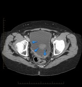
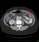
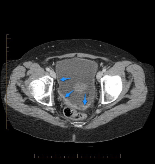
Peritoneal fluid is present which outlines multiple areas of peritoneal nodular thickening (blue arrows). In addition, the omentum to the left of the midline appears stranded with soft tissue nodules (red arrows).
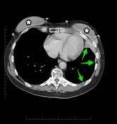
The pleura on the left has similar appearances (green arrows), and multiple mixed sclerotic/lytic lesions are seen the largest involving the right pedicle and adjacent vertebral body of T12 (yellow dotted line).
Bilateral breast prostheses are noted (*).
Case Discussion
This case illustrates typical CT appearances of widespread peritoneal (as well as pleural and bony) metastatic disease from prior breast cancer.


 Unable to process the form. Check for errors and try again.