
Presentation
Two days pleuritic chest pain, no shortness of breath. Tachycardic, normal oxygen saturations.
Patient Data
Age: 20 years
Gender: Male
From the case:
Pneumomediastinum
Download
Info
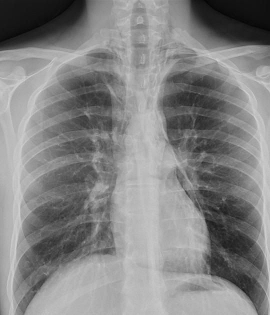
Small left apical pneumothorax.
Subcutaneous emphysema (vertical lucent stripes into neck) and "double bronchial wall sign" suggesting pneumomediastinum. Small rim of air at AP window.
Case Discussion
This is a classical appearance of pneumomediastinum.
With the imaging and the stable status of the patient, this was likely from alveolar rupture allowing air to track into the mediastinum which is a recognized cause of pneumomediastinum.
This can be managed conservatively.


 Unable to process the form. Check for errors and try again.