
Presentation
Immunocompromised status. Now bed bound.
Patient Data


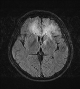
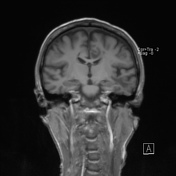

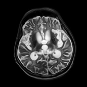
Mild cerebral atrophy in both cerebral hemispheres.
Extensive high confluent T2/FLAIR signal in the subcortical and deep white matter of both frontal lobes and the left temporal lobe.
Cerebellum and brainstem normal.

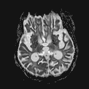

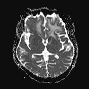
MRI 7 mth later following a deterioration in neurological status
Extensive symmetrical confluent bilateral deep white matter T2 high signal - now throughout both cerebral hemispheres, compared to only the frontal and left temporal lobes on the previous scan.
New T2 signal throughout the pons.
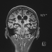
Extensive cerebral atrophy excessive for age which has progressed markedly from the scan 7 months earlier.
Cerebellar atrophy with symmetrical high T2 signal in the medial aspects of both hemispheres.
Case Discussion
Progressive multifocal leukoencephalopathy (PML) is a demyelinating disease which results from the JC virus infecting oligodendrocytes. It is considered the most common clinical manifestation of JC virus infection in the brain. Although often in those with AIDS, it can occur in any patient with an immunocompromised status.
Multifocal T2 high signal foci representing areas of demyelination are characteristic with both the subcortical and deep white matter is involved with a preponderance for the subcortical U fibers. PML has a propensity for the frontal and parieto-occipital regions as well as the thalamus and basal ganglia.
This case illustrates the rapidity (progressiveness) of the disease.


 Unable to process the form. Check for errors and try again.