
Presentation
Acute onset of intense pelvic pain with a recent history of intermenstrual bleeding.
Patient Data
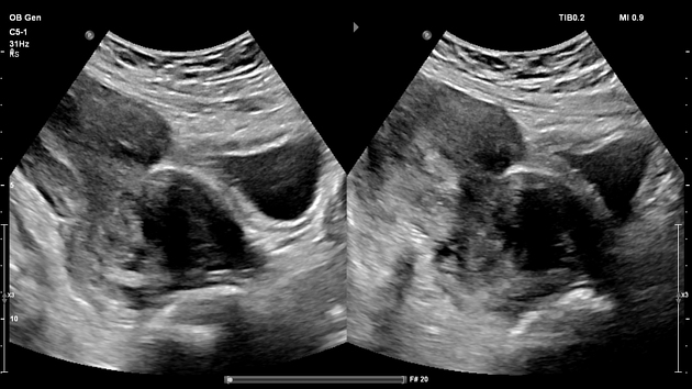
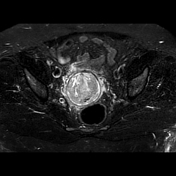
Hourglass appearance of the uterus due to cervical distention by a well-circumscribed hypoechoic solid mass measuring up to 6 cm in diameter.
Additional findings: fatty liver, gallbladder sludge with two small polyps, and a left paraumbilical anterior abdominal wall hernia with omental fat prolapse.


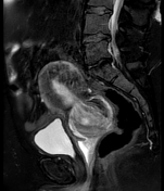
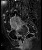


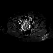

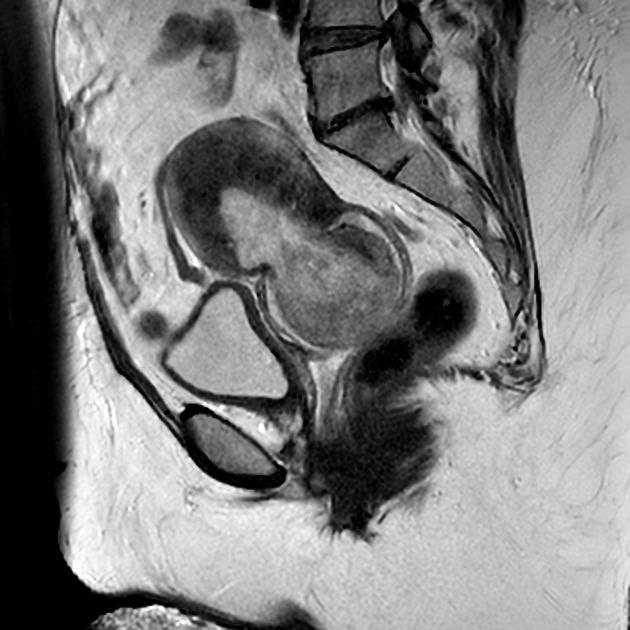
Further evaluation with standard pelvic MRI protocol 1.5 T MRI showed a round well-defined soft tissue pedunculated mass arising from the endometrium and protruding downwards into the cervical canal.
The polyp signal intensity is equal to the endometrium in all sequences except for the post-contrast study which demonstrates a complete lack of enhancement in keeping with infarction.
Note the fine linear enhancing peduncular strands at the base of the polyp radiating in a windmill pattern (best seen while scrolling the images on T1 fat sat C+ sagittal plan) consistent with a twisted pedicle.
A rim of fluid surrounds the polyp filling the vaginal fornix also.
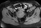
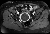
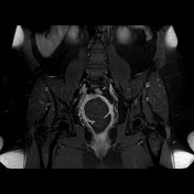
Additional findings: Diffuse muscle wall and junctional zone thickening with micronodular foci of high T1 and T2 signal intensity dispersed throughout the myometrium giving it a stratified appearance and lost endo/myometrial junction definition suggestive of diffuse adenomyosis.
Case Discussion
A case of prolapsed endometrial polyp with twisted pedicle and resultant infarction in a perimenopausal woman with coexistent adenomyosis presenting with acute onset of intense pelvic pain and a history of intermenstrual bleeding.
Endometrial polyps are benign sessile or pedunculated nodules arising from the endometrial surface and sometimes protruding into the cervical canal giving it the hourglass appearance as in this case.
They are mostly encountered in middle aged women and the incidence increases with age 1.
Clinical presentation varies between asymptomatic to infertile women with intermenstrual or postmenopausal bleeding 2.
Usually they are well-defined nodules with multimodal imaging appearance similar to the endometrial tissue (hyperechoic, hypoattenuating and with equal signal intensity to the endometrium in almost all sequences) surrounded by endometrium and a rim of fluid.
Regarding post-contrast studies the pattern of enhancement may vary between homogenous to heterogenous enhancement relative to the endometrium but rarely they appear as non-enhancing nodules in case of a twisted pedicle with resultant infarction as in this case.
Treatment strategy consists in polypectomy followed by histopathologic examination to rule out possible endometrial carcinoma within the polyp 3.


 Unable to process the form. Check for errors and try again.