
Presentation
Abdominal pain.
Patient Data


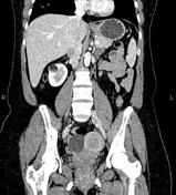
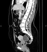
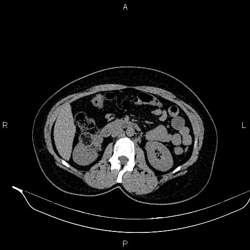
A 25 x 20 mm well-defined partially exophytic fat containing mass is noted at mid portion of right kidney with a heterogeneous enhancement on postcontrast images, inferring angiomyolipoma.
The uterus contains several fibroids less than 35 mm.
Three adjacent cystic lesions without enhancing solid component are seen posterior to the uterus in keeping with ovarian cysts, probably endometriomas.
Case Discussion
Renal angiomyolipomas are a type of benign renal neoplasms encountered both sporadically and as part of a phakomatosis, most commonly tuberous sclerosis. They usually have characteristic radiographic appearances with fat density components. Most lesions involve the cortex and demonstrate macroscopic fat (less than -20 HU).


 Unable to process the form. Check for errors and try again.