
Presentation
Incidental left renal mass observed on ultrasound examination for alternative indication.
Patient Data
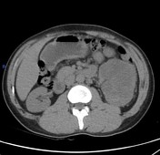

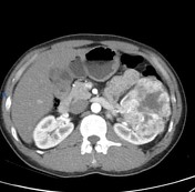
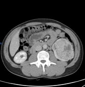
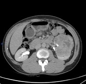
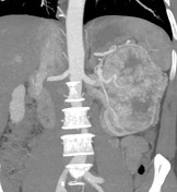
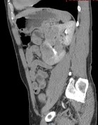
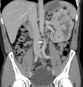

Left renal fairly defined heterogeneous soft tissue mass lesion is seen implicating mainly left renal anteromedial aspect, it measures roughly about 9.5 x 8.2 x 11.5 cm along its maximum AP, TV and CC dimensions respectively diameters , this soft tissue mass lesion is seen replacing most of anterior aspect of the kidney sparing its upper pole, and overlying left renal hilum , moreover extending anteriorly and laterally engulfing renal capsule and along perinephric planes , it compress the descending colon anterolaterally, superiorly it is seen abutting the splenic hilum,medially the mass is abutting the renal pelvis and overlying renal hilum with amputated anterior calyces, however sparing reno-vascular pedicle which appears fairly opacified with multiple feeder branches to the mass, the mass shows significant heterogeneous enhancement with multiple central non enhanced hypodense areas likely necrotic/degenerated components , no areas of hemorrhage is seen inside the mass and no evidence of extracapsular spread.
Moreover, multiple discrete para-aortic lymphadenopathies are also seen, the largest about 1.5x1.4 cm at the left para-aortic group.
There is a solitary small hepatic focal lesion seen related to the hepatic dome (segment IVa) measuring about 8 mm and appears vivid enhancement at arterial phase then fades rapidly.
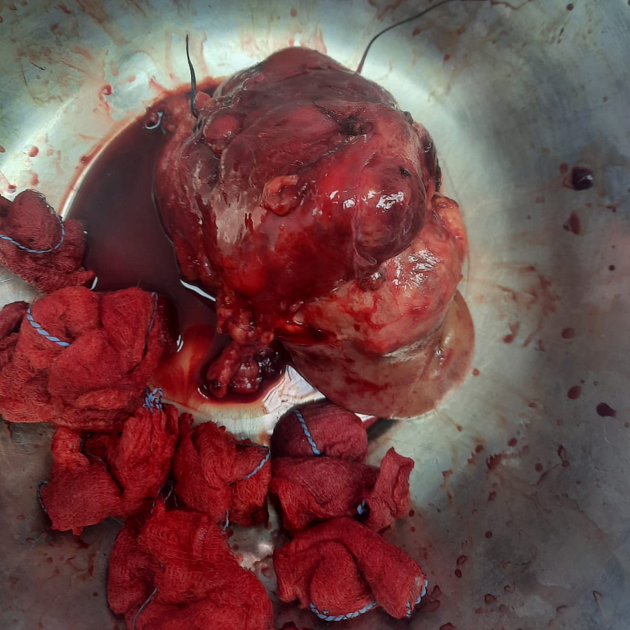
The patient underwent total nephrectomy and regional nodal resection, and then histopathology evaluation confirmed low-grade clear cell renal cell carcinoma.
This image is courtesy of Dr. Ahmed Elzanaty (urology surgeon).
Case Discussion
The findings are highly suggestive of left renal neoplastic soft tissue mass lesion with necrotic changes (associated with small para-aortic lymphadenopathy as well as a small solitary hepatic focal lesion.
The patient underwent a total nephrectomy, which confirmed the diagnosis of a renal cell carcinoma (RCC). (T2b N1 M1)
RCC is a malignant tumor derived from the renal epithelium being the most common malignant renal tumor, with a variety of radiographic appearances.


 Unable to process the form. Check for errors and try again.