
Presentation
Hematuria and right flank mass.
Patient Data
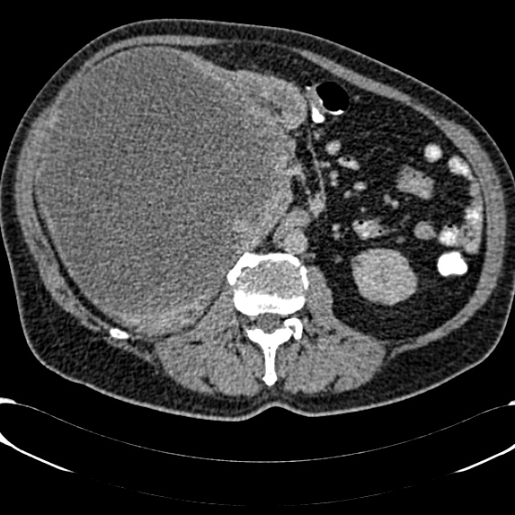
Huge heterogenousmass with necrotic elements essentially replacing the right kidney,
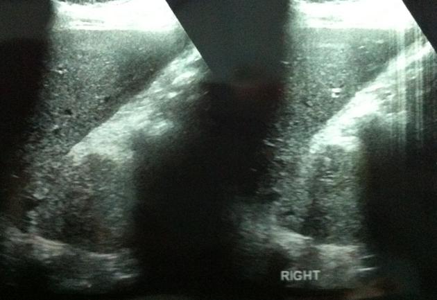
Due to renal impairment initial follow up imaging was with ultrasound.
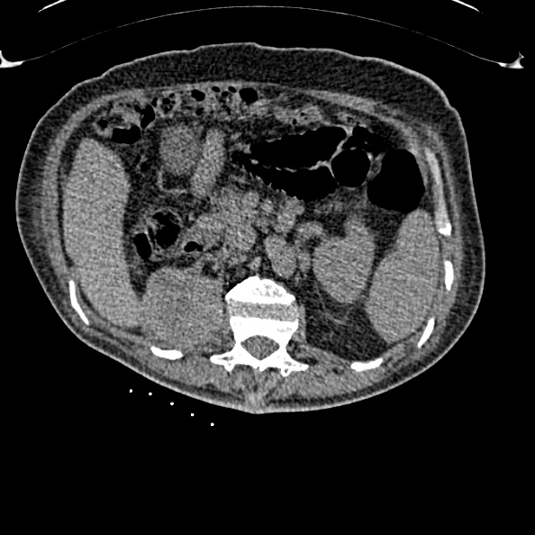
Right nephrectomy. Soft tissue mass in the renal bed.
CT imaging performed to assess further.
The clinical team requested histological confirmation of recurrence, with an image guided biopsy.
The soft tissue recurrence in the renal bed is confirmed as well as multiple destructive bony metastases.
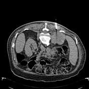

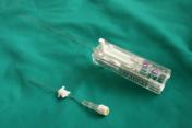

Prone position. 18G co-axial needle core biopsy performed under ultrasound guidance. Ultrasound guided biopsy could also have been performed, but difficulties with positioning due to the associated spinal metastases, made CT guidance more straightforward.
Case Discussion
Renal cell carcinoma (RCC) is the commonest renal malignancy. It classically presents with the triad of haemautria, pain and a flank mass, but in contemporary medicine this is rarely the case. in fact, more than 50% of renal cell carcinomas are now identified on imaging investigations performed for other purposes.
Renal cell carcinoma is usually avidly enhancing on contrast enhanced CT, as are its metastases. In addition to locoregional nodes, the liver. lungs and bones are common sites for metastases. RCC is one of the tumors causing cannonball metastases to the lungs and bone metastases are lytic and destructive. Renal cell carcinoma is the common primary to metastasize to the pancreas.


 Unable to process the form. Check for errors and try again.