
Presentation
Evaluation of pacemaker in patient who has a history of a bicuspid aortic valve who underwent a Ross procedure in past.
Patient Data
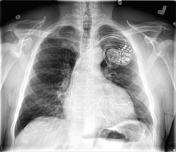
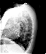

The heart is borderline enlarged. Aortic root and arch ( pulmonary homograft ) is diffusely calcified.
Sternal wires are demonstrated, which are intact.
A left-sided triple lead pacemaker is demonstrated with leads in the right atrium, right ventricular apex and posterior left ventricular wall via coronary sinus. No pneumothorax, mediastinal widening, CHF, or pleural fluid demonstrated.
Case Discussion
Echo cardiogram
Aortic Valve:
Mild AI. Mean PG = 5 mmHg. Ross procedure.
Mitral Valve:
The mitral valve appears normal. There is no evidence of mitral stenosis. There is mild to moderate mitral regurgitation.
Tricuspid Valve:
The tricuspid valve is normal. There is no evidence of tricuspid stenosis. There is mild tricuspid regurgitation. There is no evidence of pulmonary hypertension. The Right Ventricular Systolic Pressure is calculated at 34 mmHg.
Pulmonic Valve:
The pulmonic valve is not well visualized. There is a bioprosthetic pulmonic valve. Mild PI, Mean gradient 9 mmHg.


 Unable to process the form. Check for errors and try again.