
Presentation
Abdominal pain
Patient Data

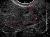

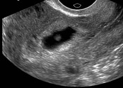
The initial study showed what appeared to be a possible gestational sac in the lower segment of the uterus. The radiologist recommended follow up and further Beta HCG correlation.
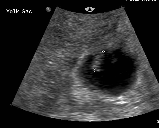
If you look carefully at the images you will see the gestational sac is developing in the myometrium. The presumed gestational sac is larger on ultrasound and a yolk sac is present. The patient had further elevation of her Beta HCG levels. Her symptoms persisted.
On the very early images the endometrial stripe is visible passing posterior to the developing gestational sac.
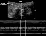

At the third study 12 days later a fetal pole was visible. M-mode tracing confirmed a fetal heartbeat.
Case Discussion
A gravida 2 presents to the ED with lower abdominal pain. Previous C-section delivery. The patient has an elevated Beta HCG level. She is referred for imaging with a provisional diagnosis of ? ectopic pregnancy. This turned out to be an ectopic pregnancy in the surgical scar of a previous cesarian section. On real time imaging you could see the scar in the lower segment of the uterus and clearly visualize the sac in the scar itself. The pregnancy was terminated chemically.


 Unable to process the form. Check for errors and try again.