
Presentation
Multiple episodes of melena. Iron deficiency anemia.
Patient Data
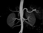

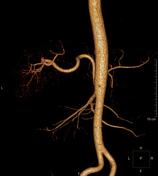
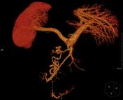
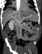


Focal small nodular areas of enhancement and bulbous swelling of the intramural veins in the wall of the jejunum, undetectable on arterial phase and brightest on enteric phase. Enlarged draining antimesenteric jejunal veins.
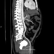
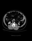
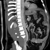
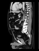
Mesenteric congestion. The jejunal bowel loops show circumferential mural thickening.

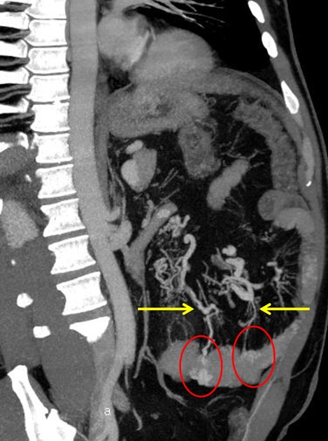
The first image shows focal small nodular areas of enhancement and bulbous swelling of the intramural veins in the wall of the jejunum (red circles), undetectable on arterial phase and brightest on enteric phase. Enlarged draining antimesenteric jejunal veins (yellow arrows).
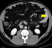
The second image shows marker (chevron) at the site of the ablated jejunal lesions using balloon-assisted endoscopy. Mesenteric smudging (congestion) is also noted.
Case Discussion
This patient had a balloon-assisted endoscopy which confirmed the jejunal angioectasia and the lesions were ablated with a marker applied to the site of intervention. Congestive enteropathy was also noted at endoscopy.
Angioectasia is the most common cause of obscure gastrointestinal bleeding.
they consist of thin tortuous veins that lack an internal elastic layer
peak incidence: 7th and 8th decades of life
Endoscopy: angioectasias consist of punctate or patchy areas of erythema, 2–10 mm in size.
CT: focal punctate or discoid areas of enhancement <5 mm in size or bulbous swelling of the intramural vessels in the wall of the small bowel, especially in the jejunum. Enhancement is brightest during the enteric phase and fades somewhat during the delayed phase. These lesions don not enhance during the arterial phase, which distinguishes them from arterial lesions.


 Unable to process the form. Check for errors and try again.