Presentation
On and off mid back pain since 2 years.
Patient Data


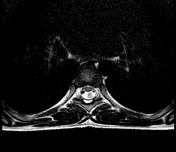

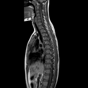

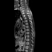

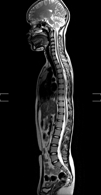
There is a well-defined CSF signal intensity non-enhancing lesion in the posterior intradural extramedullary region from T4-6 level, causing mild thinning and anteriorly displacing the spinal cord measuring 35 x 8 mm. Multiple rounded T2 hypointense foci without post-contrast enhancement noted above and below the above-mentioned lesion, suggestive of CSF flow artifacts. The spinal cord reveals normal signal intensity. Findings are suggestive of thoracic arachnoid cyst from T4-6 level.
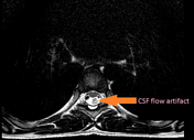
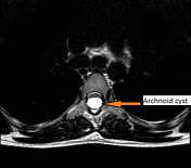
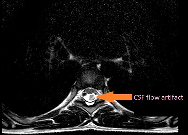
Arrows point to CSF pulsation artifact and the thoracic arachnoid cyst.
Case Discussion
Patient had complaints of on and off mid back pain for 2 years. No history of trauma, tingling or numbness in upper and lower limbs. Previous radiograph was unremarkable, hence further evaluation with MRI was advised. After MRI diagnosis of thoracic arachnoid cyst, patient was operated and was advised follow-up imaging after 3 months.


 Unable to process the form. Check for errors and try again.