
Presentation
Recent unintentional weight loss.
Patient Data


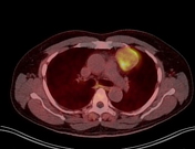
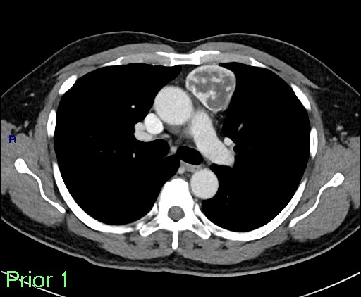
5 cm sharply demarcated, mildly lobulated triangular prevascular mediastinal mass with central cystic/necrotic change and enhancing solid components. Low-level avidity on PET (SUVmax 4.2).
Normal lymph nodes and pleura. No metastases.
Incidental enlarged prostate and uncomplicated sigmoid diverticulosis.
Case Discussion
Histology demonstrated type AB thymoma and the tumor is Masaoka stage 1, with no capsular invasion or spread elsewhere. Thymic epithelial tumors can be DOTATATE-avid and this can be used as an alternative means of disease control 1.
The mass was surgically resected and the report showed:
"Specimen Type:
A: (1) MEDIASTINAL MASS
B: (2) THYMUS (RIGHT SIDE)
C: (3) LEFT SIDE THYMUS
Clinical Details: mediastinal mass and thymus.
Macroscopic Description:
A. (1) MEDIASTINAL MASS:
Pots, forms, cassettes, and co-paths are matching. Part a contains a smooth round piece of pink tissue which measures 49 x 43 x 36 mm and which is un-oriented. It contains several metallic clips on the surface, possibly representing nerve margins. The specimen weighs 52 g. On slicing the mass comprises an encapsulated and partly cystic lesion with thin colourless fluid and occasional areas of hemorrhage.
Block key
A1: Possible nerve margins
A2A7: Sections through mass
Tissue remaining
DN
B. (2) THYMUS (RIGHT SIDE):
Pot B contains a single piece of un-oriented fatty tissue which measures 111 mm x 47 mm x 23 mm. The specimen weighs 40 g. Prior to slicing it is inked green on the external surface. On slicing the specimen appears to comprise soft fat only with no lesions.
Block key
B1B2 sections through specimen
Tissue remaining
DN.
C. (3) LEFT SIDE THYMUS:
Pot C contains a single piece of ragged fatty and un-oriented tissue. It measures 65 x 43 x 28 mm. The specimen weighs 40.5 g. Prior to slicing the external surface is inked with green ink. On slicing the specimen appears to comprise only soft fat with no lesions.
Block key
C1-C2: Representative sections
Tissue remains
DN.
Microscopic Description:
A. (1) MEDIASTINAL MASS:
previous treatment (neoadjuvant chemotherapy and/or radiotherapy): No
specimen type: Thymus plus surrounding tissue (radical thymectomy)
MACROSCOPIC FEATURES
location of tumor (intrathymic)
tumor size: 49 mm (maximum dimension)
MICROSCOPIC FEATURES
Histological type: Thymoma AB Type A(90%); type B (type B1):10%. There are widespread cystic areas. No nerve is identified microscopically.
Direct Invasion
capsule: No invasion beyond capsule
mediastinal Pleura: Not applicable
pericardium: Not applicable
Separate extra thymic tumor nodules: None
Lymph node involvement: No lymph nodes identified.
Margins
excision complete (R0): Yes
closest margin <1 mm
B. (2) THYMUS (RIGHT SIDE): Adipose tissue showing microscopic calcification and a single unremarkable lymph node.
C. (3) LEFT SIDE THYMUS: Unremarkable thymic tissue.
Final Diagnosis:
ANTERIOR MEDIASTINAL MASS/ THYMECTOMY:
Histological type: Thymoma, type AB
Lymph nodes: 0/1
TNM Stage (8th Edition): Ia N0 R0


 Unable to process the form. Check for errors and try again.