
Presentation
Tetralogy of Fallot. Surgically-corrected VSD and bioprosthetic pulmonary valve. Presented with shortness of breath. Underwent a second pulmonary valve replacement.
Patient Data
Age: 20 years
Gender: Male

Download
Info

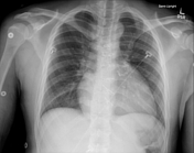
CXR 1
Original Carpentier-Edwards valve. Sternotomy wires of two kinds.
Ventricular septal defect (VSD) repair first.
Bioprosthetic valve in a right ventricular outflow tract (RVOT) conduit.
CXR 2
Postoperative image showing the percutaneously placed pulmonary valve on top of the older valve.
Case Discussion
Conclusions of cardiac MRI prior to transcatheter pulmonary valve replacement:
- patient born with tetralogy of Fallot. Surgically corrected VSD and prosthetic pulmonary valve are seen. 5-vessel aortic arch with a retroesophageal course of the right subclavian artery arising from the descending thoracic aorta.
- RVOT measures 38 mm in its maximal dimension. The main pulmonary artery measures 24 mm in maximal dimension. Right and left pulmonary arteries measure 21 and 24 mm in diameter.
- right ventricle: right ventricular dilatation, right ventricular hypertrophy, RV end diastolic volume index at least 135 mL. RV ejection fraction 53%.
- valvular function: severe pulmonary regurgitation of the prosthetic pulmonary valve (57%).
- delayed enhancement: none seen.
Therefore a transcatheter valve has been placed due to evidence of failure of the original valve.


 Unable to process the form. Check for errors and try again.