
Presentation
Hematuria.
Patient Data
Age: 2 years
Gender: Male
From the case:
Wilms tumor


Download
Info
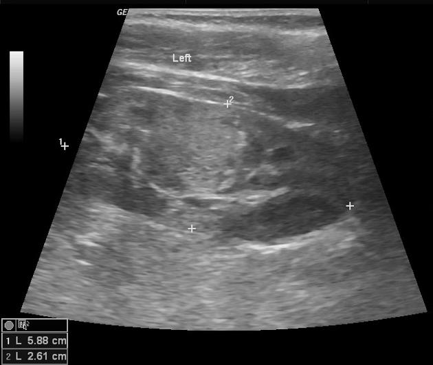
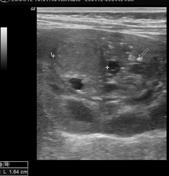
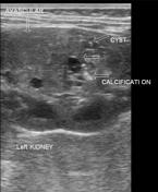
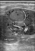
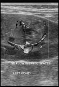
LEFT kidney shows a heterogeneous lesion (26 mm maximum dimention) in interpolar region.
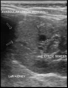
Cranially there is an avascular rounded lesion (18 x 12 x 11 mm) with thin wall. Vascularity is present in the wall only. No central vascularity is noted.
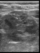
In the caudal part, there are multiple tiny cystic spaces with tiny echogenic foci without posterior acoustic shadowing -- possibly tiny calcifications. No vascularity is noted in this caudal part of lesion.
Case Discussion
Left nephrectomy performed, revealing Wilms tumor.


 Unable to process the form. Check for errors and try again.